5LGR | pdb_00005lgr

Crystal structure of mouse CARM1 in complex with ligand P1C3u


Experimental Data Snapshot

  • Method: X-RAY DIFFRACTION
  • Resolution: 2.00 Å
  • R-Value Free: 
    0.234 (Depositor), 0.234 (DCC) 
  • R-Value Work: 
    0.192 (Depositor), 0.195 (DCC) 
  • R-Value Observed: 
    0.194 (Depositor) 

Starting Model: experimental
View more details

wwPDB Validation 3D Report Full Report

Validation slider image for 5LGR

Ligand Structure Quality Assessment 


This is version 1.3 of the entry. See complete history

Literature

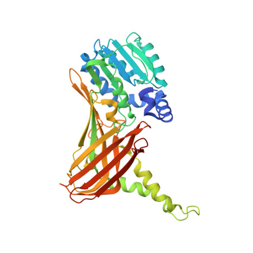
Transition state mimics are valuable mechanistic probes for structural studies with the arginine methyltransferase CARM1.

van Haren, M.J.Marechal, N.Troffer-Charlier, N.Cianciulli, A.Sbardella, G.Cavarelli, J.Martin, N.I.

(2017) Proc Natl Acad Sci U S A 114: 3625-3630

  • DOI: https://doi.org/10.1073/pnas.1618401114
  • Primary Citation Related Structures: 
    5LGP, 5LGQ, 5LGR, 5LGS

  • PubMed Abstract: 

    Coactivator associated arginine methyltransferase 1 (CARM1) is a member of the protein arginine methyltransferase (PRMT) family and methylates a range of proteins in eukaryotic cells. Overexpression of CARM1 is implicated in a number of cancers, and it is therefore seen as a potential therapeutic target. Peptide sequences derived from the well-defined CARM1 substrate poly(A)-binding protein 1 (PABP1) were covalently linked to an adenosine moiety as in the AdoMet cofactor to generate transition state mimics. These constructs were found to be potent CARM1 inhibitors and also formed stable complexes with the enzyme. High-resolution crystal structures of CARM1 in complex with these compounds confirm a mode of binding that is indeed reflective of the transition state at the CARM1 active site. Given the transient nature of PRMT-substrate complexes, such transition state mimics represent valuable chemical tools for structural studies aimed at deciphering the regulation of arginine methylation mediated by the family of arginine methyltransferases.


  • Organizational Affiliation
    • Department of Chemical Biology & Drug Discovery, Utrecht Institute for Pharmaceutical Sciences, Utrecht University, 3584 CG Utrecht, The Netherlands.

Macromolecule Content 

  • Total Structure Weight: 171.08 kDa 
  • Atom Count: 12,311 
  • Modeled Residue Count: 1,417 
  • Deposited Residue Count: 1,492 
  • Unique protein chains: 2

Macromolecules

Find similar proteins by:|  3D Structure
Entity ID: 1
MoleculeChains  Sequence LengthOrganismDetailsImage
Histone-arginine methyltransferase CARM1
A, B, C, D
361Mus musculusMutation(s): 0 
Gene Names: Carm1Prmt4
EC: 2.1.1.319
UniProt & NIH Common Fund Data Resources
Find proteins for Q9WVG6 (Mus musculus)
Explore Q9WVG6 
Go to UniProtKB:  Q9WVG6
IMPC:  MGI:1913208
Entity Groups
Sequence Clusters30% Identity50% Identity70% Identity90% Identity95% Identity100% Identity
UniProt GroupQ9WVG6
Sequence Annotations
Expand
Reference Sequence
Find similar proteins by:  Sequence   |   3D Structure  
Entity ID: 2
MoleculeChains  Sequence LengthOrganismDetailsImage
Polyadenylate-binding protein 1
E, F, G, H
12Homo sapiensMutation(s): 0 
UniProt & NIH Common Fund Data Resources
Find proteins for P11940 (Homo sapiens)
Explore P11940 
Go to UniProtKB:  P11940
PHAROS:  P11940
GTEx:  ENSG00000070756 
Entity Groups
Sequence Clusters30% Identity50% Identity70% Identity90% Identity95% Identity100% Identity
UniProt GroupP11940
Sequence Annotations
Expand
Reference Sequence

Small Molecules

Ligands 8 Unique
IDChains Name / Formula / InChI Key2D Diagram3D Interactions
QVR

Query on QVR


Download:Ideal Coordinates CCD File
AA [auth H],
R [auth E],
V [auth F],
Y [auth G]
(2~{R},3~{R},4~{S},5~{R})-2-(6-aminopurin-9-yl)-5-[(~{E})-prop-1-enyl]oxolane-3,4-diol
C12 H15 N5 O3
UYHMWDPUDJRZGB-JVINVVEESA-N
PG6

Query on PG6


Download:Ideal Coordinates CCD File
N [auth B]1-(2-METHOXY-ETHOXY)-2-{2-[2-(2-METHOXY-ETHOXY]-ETHOXY}-ETHANE
C12 H26 O6
DMDPGPKXQDIQQG-UHFFFAOYSA-N
LPD

Query on LPD


Download:Ideal Coordinates CCD File
BA [auth H],
S [auth E],
W [auth F],
X [auth G]
L-PROLINAMIDE
C5 H10 N2 O
VLJNHYLEOZPXFW-BYPYZUCNSA-N
PEG

Query on PEG


Download:Ideal Coordinates CCD File
P [auth C]DI(HYDROXYETHYL)ETHER
C4 H10 O3
MTHSVFCYNBDYFN-UHFFFAOYSA-N
SO4

Query on SO4


Download:Ideal Coordinates CCD File
I [auth A]SULFATE ION
O4 S
QAOWNCQODCNURD-UHFFFAOYSA-L
DXE

Query on DXE


Download:Ideal Coordinates CCD File
L [auth A]1,2-DIMETHOXYETHANE
C4 H10 O2
XTHFKEDIFFGKHM-UHFFFAOYSA-N
EDO

Query on EDO


Download:Ideal Coordinates CCD File
J [auth A]
K [auth A]
M [auth B]
O [auth C]
Q [auth D]
J [auth A],
K [auth A],
M [auth B],
O [auth C],
Q [auth D],
T [auth E]
1,2-ETHANEDIOL
C2 H6 O2
LYCAIKOWRPUZTN-UHFFFAOYSA-N
ACE

Query on ACE


Download:Ideal Coordinates CCD File
U [auth F],
Z [auth H]
ACETYL GROUP
C2 H4 O
IKHGUXGNUITLKF-UHFFFAOYSA-N

Experimental Data & Validation

Experimental Data

  • Method: X-RAY DIFFRACTION
  • Resolution: 2.00 Å
  • R-Value Free:  0.234 (Depositor), 0.234 (DCC) 
  • R-Value Work:  0.192 (Depositor), 0.195 (DCC) 
  • R-Value Observed: 0.194 (Depositor) 
Space Group: P 21 21 2
Unit Cell:
Length ( Å )Angle ( ˚ )
a = 74.831α = 90
b = 98.544β = 90
c = 208.336γ = 90
Software Package:
Software NamePurpose
XDSdata reduction
Aimlessdata scaling
PHENIXrefinement
PDB_EXTRACTdata extraction

Structure Validation

View Full Validation Report


Ligand Structure Quality Assessment 


Entry History 

Deposition Data

Revision History  (Full details and data files)

  • Version 1.0: 2017-03-22
    Type: Initial release
  • Version 1.1: 2017-04-05
    Changes: Database references
  • Version 1.2: 2017-04-12
    Changes: Database references
  • Version 1.3: 2024-01-10
    Changes: Data collection, Database references, Refinement description