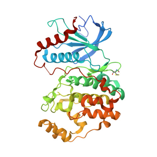
5LCJ

In-Gel Activity-Based Protein Profiling of a Clickable Covalent Erk 1/2 Inhibitor


Experimental Data Snapshot

  • Method: X-RAY DIFFRACTION
  • Resolution: 1.78 Å
  • R-Value Free: 0.212 
  • R-Value Work: 0.162 
  • R-Value Observed: 0.165 

wwPDB Validation   3D Report Full Report


Ligand Structure Quality Assessment 


This is version 1.1 of the entry. See complete history


Literature

In-gel activity-based protein profiling of a clickable covalent ERK1/2 inhibitor.

Lebraud, H.Wright, D.J.East, C.E.Holding, F.P.O'Reilly, M.Heightman, T.D.

(2016) Mol Biosyst 12: 2867-2874

  • DOI: https://doi.org/10.1039/c6mb00367b
  • Primary Citation of Related Structures:  
    5LCJ, 5LCK

  • PubMed Abstract: 

    In-gel activity-based protein profiling (ABPP) offers rapid assessment of the proteome-wide selectivity and target engagement of a chemical tool. Here we demonstrate the use of the inverse electron demand Diels Alder (IEDDA) click reaction for in-gel ABPP by evaluating the selectivity profile and target engagement of a covalent ERK1/2 probe tagged with a trans-cyclooctene group. The chemical probe was shown to bind covalently to Cys166 of ERK2 using protein MS and X-ray crystallography, and displayed submicromolar GI50s in A375 and HCT116 cells. In both cell lines, the probe demonstrated target engagement and a good selectivity profile at low concentrations, which was lost at higher concentrations. The IEDDA cycloaddition enabled fast and quantitative fluorescent tagging for readout with a high background-to-noise ratio and thereby provides a promising alternative to the commonly used copper catalysed alkyne-azide cycloaddition.


  • Organizational Affiliation

    Astex Pharmaceuticals, 436 Cambridge Science Park, Cambridge, CB4 0QA, UK. Tom.Heightman@astx.com.


Macromolecules
Find similar proteins by:  (by identity cutoff)  |  3D Structure
Entity ID: 1
MoleculeChains Sequence LengthOrganismDetailsImage
Mitogen-activated protein kinase 1368Homo sapiensMutation(s): 0 
Gene Names: MAPK1ERK2PRKM1PRKM2
EC: 2.7.11.24
UniProt & NIH Common Fund Data Resources
Find proteins for P28482 (Homo sapiens)
Explore P28482 
Go to UniProtKB:  P28482
PHAROS:  P28482
GTEx:  ENSG00000100030 
Entity Groups  
Sequence Clusters30% Identity50% Identity70% Identity90% Identity95% Identity100% Identity
UniProt GroupP28482
Sequence Annotations
Expand
  • Reference Sequence
Small Molecules
Ligands 2 Unique
IDChains Name / Formula / InChI Key2D Diagram3D Interactions
6TS
Query on 6TS

Download Ideal Coordinates CCD File 
B [auth A][(1~{R},4~{Z})-cyclooct-4-en-1-yl] ~{N}-[4-[4-[[5-chloranyl-4-[[2-(propanoylamino)phenyl]amino]pyrimidin-2-yl]amino]pyridin-2-yl]but-3-ynyl]carbamate
C31 H34 Cl N7 O3
DLZIOTBNYZNPPK-YJMIJIGASA-N
SO4
Query on SO4

Download Ideal Coordinates CCD File 
C [auth A],
D [auth A]
SULFATE ION
O4 S
QAOWNCQODCNURD-UHFFFAOYSA-L
Modified Residues  1 Unique
IDChains TypeFormula2D DiagramParent
CME
Query on CME
A
L-PEPTIDE LINKINGC5 H11 N O3 S2CYS
Experimental Data & Validation

Experimental Data

  • Method: X-RAY DIFFRACTION
  • Resolution: 1.78 Å
  • R-Value Free: 0.212 
  • R-Value Work: 0.162 
  • R-Value Observed: 0.165 
  • Space Group: P 1 21 1
Unit Cell:
Length ( Å )Angle ( ˚ )
a = 48.729α = 90
b = 70.807β = 109.18
c = 60.208γ = 90
Software Package:
Software NamePurpose
BUSTERrefinement
iMOSFLMdata reduction
SCALEPACKdata scaling
REFMACphasing

Structure Validation

View Full Validation Report


Ligand Structure Quality Assessment 


Entry History 

Deposition Data

Revision History  (Full details and data files)

  • Version 1.0: 2016-07-20
    Type: Initial release
  • Version 1.1: 2016-08-31
    Changes: Database references