5LB7 | pdb_00005lb7

Complex structure between p60N/p80C katanin and a peptide derived from ASPM


Experimental Data Snapshot

  • Method: X-RAY DIFFRACTION
  • Resolution: 1.50 Å
  • R-Value Free: 
    0.223 (Depositor), 0.228 (DCC) 
  • R-Value Work: 
    0.183 (Depositor), 0.188 (DCC) 
  • R-Value Observed: 
    0.184 (Depositor) 

wwPDB Validation 3D Report Full Report

Validation slider image for 5LB7

This is version 1.4 of the entry. See complete history

Literature

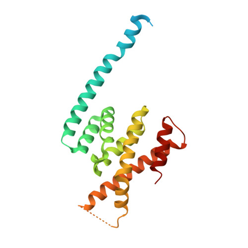
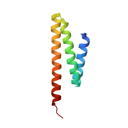
Microtubule minus-end regulation at spindle poles by an ASPM-katanin complex.

Jiang, K.Rezabkova, L.Hua, S.Liu, Q.Capitani, G.Maarten Altelaar, A.F.Heck, A.J.R.Kammerer, R.A.Steinmetz, M.O.Akhmanova, A.

(2017) Nat Cell Biol 19: 480-492

  • DOI: https://doi.org/10.1038/ncb3511
  • Primary Citation Related Structures: 
    5LB7, 5NBT

  • PubMed Abstract: 

    ASPM (known as Asp in fly and ASPM-1 in worm) is a microcephaly-associated protein family that regulates spindle architecture, but the underlying mechanism is poorly understood. Here, we show that ASPM forms a complex with another protein linked to microcephaly, the microtubule-severing ATPase katanin. ASPM and katanin localize to spindle poles in a mutually dependent manner and regulate spindle flux. X-ray crystallography revealed that the heterodimer formed by the N- and C-terminal domains of the katanin subunits p60 and p80, respectively, binds conserved motifs in ASPM. Reconstitution experiments demonstrated that ASPM autonomously tracks growing microtubule minus ends and inhibits their growth, while katanin decorates and bends both ends of dynamic microtubules and potentiates the minus-end blocking activity of ASPM. ASPM also binds along microtubules, recruits katanin and promotes katanin-mediated severing of dynamic microtubules. We propose that the ASPM-katanin complex controls microtubule disassembly at spindle poles and that misregulation of this process can lead to microcephaly.


  • Organizational Affiliation
    • Cell Biology, Department of Biology, Faculty of Science, Utrecht University, Padualaan 8, 3584 CH Utrecht, The Netherlands.

Macromolecule Content 

  • Total Structure Weight: 33.89 kDa 
  • Atom Count: 2,216 
  • Modeled Residue Count: 247 
  • Deposited Residue Count: 301 
  • Unique protein chains: 3

Macromolecules

Find similar proteins by:|  3D Structure
Entity ID: 1
MoleculeChains  Sequence LengthOrganismDetailsImage
Katanin p80 WD40 repeat-containing subunit B1212Mus musculusMutation(s): 0 
Gene Names: Katnb1
UniProt & NIH Common Fund Data Resources
Find proteins for Q8BG40 (Mus musculus)
Explore Q8BG40 
Go to UniProtKB:  Q8BG40
IMPC:  MGI:1921437
Entity Groups
Sequence Clusters30% Identity50% Identity70% Identity90% Identity95% Identity100% Identity
UniProt GroupQ8BG40
Sequence Annotations
Expand
Reference Sequence
Find similar proteins by:|  3D Structure
Entity ID: 2
MoleculeChains  Sequence LengthOrganismDetailsImage
Katanin p60 ATPase-containing subunit A180Mus musculusMutation(s): 0 
Gene Names: Katna1
EC: 3.6.4.3 (PDB Primary Data), 5.6.1.1 (UniProt)
UniProt & NIH Common Fund Data Resources
Find proteins for Q9WV86 (Mus musculus)
Explore Q9WV86 
Go to UniProtKB:  Q9WV86
IMPC:  MGI:1344353
Entity Groups
Sequence Clusters30% Identity50% Identity70% Identity90% Identity95% Identity100% Identity
UniProt GroupQ9WV86
Sequence Annotations
Expand
Reference Sequence
Find similar proteins by:  Sequence   |   3D Structure  
Entity ID: 3
MoleculeChains  Sequence LengthOrganismDetailsImage
Abnormal spindle-like microcephaly-associated protein homolog9Mus musculusMutation(s): 0 
UniProt & NIH Common Fund Data Resources
Find proteins for Q8CJ27 (Mus musculus)
Explore Q8CJ27 
Go to UniProtKB:  Q8CJ27
IMPC:  MGI:1334448
Entity Groups
UniProt GroupQ8CJ27
Sequence Annotations
Expand
Reference Sequence

Experimental Data & Validation

Experimental Data

  • Method: X-RAY DIFFRACTION
  • Resolution: 1.50 Å
  • R-Value Free:  0.223 (Depositor), 0.228 (DCC) 
  • R-Value Work:  0.183 (Depositor), 0.188 (DCC) 
  • R-Value Observed: 0.184 (Depositor) 
Space Group: C 2 2 2
Unit Cell:
Length ( Å )Angle ( ˚ )
a = 104.09α = 90
b = 145.12β = 90
c = 37.56γ = 90
Software Package:
Software NamePurpose
PHENIXrefinement
XDSdata reduction
XSCALEdata scaling
PHASERphasing

Structure Validation

View Full Validation Report


Entry History 

Deposition Data

Revision History  (Full details and data files)

  • Version 1.0: 2017-04-26
    Type: Initial release
  • Version 1.1: 2017-05-03
    Changes: Database references
  • Version 1.2: 2017-05-10
    Changes: Database references
  • Version 1.3: 2019-10-16
    Changes: Data collection
  • Version 1.4: 2024-05-08
    Changes: Advisory, Data collection, Database references