5LB5 | pdb_00005lb5

Crystal structure of human RECQL5 helicase in complex with ADP/Mg (tricilinc form).


Experimental Data Snapshot

  • Method: X-RAY DIFFRACTION
  • Resolution: 2.00 Å
  • R-Value Free: 
    0.282 (Depositor), 0.298 (DCC) 
  • R-Value Work: 
    0.244 (Depositor), 0.258 (DCC) 
  • R-Value Observed: 
    0.246 (Depositor) 

Starting Model: experimental
View more details

wwPDB Validation 3D Report Full Report

Validation slider image for 5LB5

Ligand Structure Quality Assessment 


This is version 1.6 of the entry. See complete history

Literature

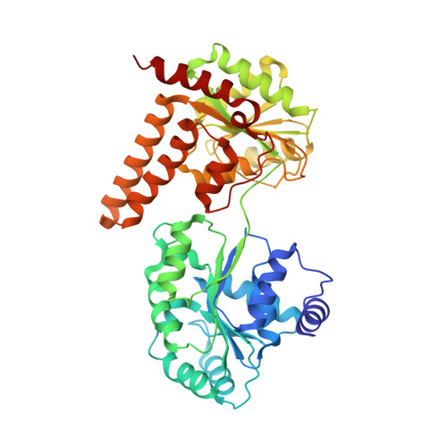
Insights into the RecQ helicase mechanism revealed by the structure of the helicase domain of human RECQL5.

Newman, J.A.Aitkenhead, H.Savitsky, P.Gileadi, O.

(2017) Nucleic Acids Res 45: 4231-4243

  • DOI: https://doi.org/10.1093/nar/gkw1362
  • Primary Citation Related Structures: 
    5LB3, 5LB5, 5LB8

  • PubMed Abstract: 

    RecQ helicases are important maintainers of genome integrity with distinct roles in almost every cellular process requiring access to DNA. RECQL5 is one of five human RecQ proteins and is particularly versatile in this regard, forming protein complexes with a diverse set of cellular partners in order to coordinate its helicase activity to various processes including replication, recombination and DNA repair. In this study, we have determined crystal structures of the core helicase domain of RECQL5 both with and without the nucleotide ADP in two distinctly different ('Open' and 'Closed') conformations. Small angle X-ray scattering studies show that the 'Open' form of the protein predominates in solution and we discuss implications of this with regards to the RECQL5 mechanism and conformational changes. We have measured the ATPase, helicase and DNA binding properties of various RECQL5 constructs and variants and discuss the role of these regions and residues in the various RECQL5 activities. Finally, we have performed a systematic comparison of the RECQL5 structures with other RecQ family structures and based on these comparisons we have constructed a model for the mechano-chemical cycle of the common catalytic core of these helicases.


  • Organizational Affiliation
    • Structural Genomics Consortium, University of Oxford, ORCRB, Roosevelt Drive, Oxford OX3 7DQ, UK.

Macromolecule Content 

  • Total Structure Weight: 200.45 kDa 
  • Atom Count: 13,859 
  • Modeled Residue Count: 1,723 
  • Deposited Residue Count: 1,780 
  • Unique protein chains: 1

Macromolecules

Find similar proteins by:|  3D Structure
Entity ID: 1
MoleculeChains  Sequence LengthOrganismDetailsImage
ATP-dependent DNA helicase Q5
A, B, C, D
445Homo sapiensMutation(s): 0 
Gene Names: RECQL5RECQ5
EC: 3.6.4.12 (PDB Primary Data), 5.6.2.4 (UniProt)
UniProt & NIH Common Fund Data Resources
Find proteins for O94762 (Homo sapiens)
Explore O94762 
Go to UniProtKB:  O94762
PHAROS:  O94762
GTEx:  ENSG00000108469 
Entity Groups
Sequence Clusters30% Identity50% Identity70% Identity90% Identity95% Identity100% Identity
UniProt GroupO94762
Sequence Annotations
Expand
Reference Sequence

Small Molecules

Ligands 4 Unique
IDChains Name / Formula / InChI Key2D Diagram3D Interactions
ADP

Query on ADP


Download:Ideal Coordinates CCD File
H [auth A],
L [auth B],
O [auth C],
S [auth D]
ADENOSINE-5'-DIPHOSPHATE
C10 H15 N5 O10 P2
XTWYTFMLZFPYCI-KQYNXXCUSA-N
DMS

Query on DMS


Download:Ideal Coordinates CCD File
F [auth A],
J [auth B],
Q [auth D]
DIMETHYL SULFOXIDE
C2 H6 O S
IAZDPXIOMUYVGZ-UHFFFAOYSA-N
ZN

Query on ZN


Download:Ideal Coordinates CCD File
E [auth A],
I [auth B],
M [auth C],
P [auth D]
ZINC ION
Zn
PTFCDOFLOPIGGS-UHFFFAOYSA-N
MG

Query on MG


Download:Ideal Coordinates CCD File
G [auth A],
K [auth B],
N [auth C],
R [auth D]
MAGNESIUM ION
Mg
JLVVSXFLKOJNIY-UHFFFAOYSA-N

Experimental Data & Validation

Experimental Data

  • Method: X-RAY DIFFRACTION
  • Resolution: 2.00 Å
  • R-Value Free:  0.282 (Depositor), 0.298 (DCC) 
  • R-Value Work:  0.244 (Depositor), 0.258 (DCC) 
  • R-Value Observed: 0.246 (Depositor) 
Space Group: P 1
Unit Cell:
Length ( Å )Angle ( ˚ )
a = 63.59α = 108.82
b = 84.75β = 90.06
c = 106.691γ = 96.86
Software Package:
Software NamePurpose
BUSTERrefinement
XDSdata reduction
XDSdata scaling
PHASERphasing

Structure Validation

View Full Validation Report


Ligand Structure Quality Assessment 


Entry History 

& Funding Information

Deposition Data


Funding OrganizationLocationGrant Number
Wellcome TrustUnited Kingdom--

Revision History  (Full details and data files)

  • Version 1.0: 2016-07-06
    Type: Initial release
  • Version 1.1: 2017-01-18
    Changes: Database references
  • Version 1.2: 2017-02-01
    Changes: Database references
  • Version 1.3: 2017-05-03
    Changes: Database references
  • Version 1.4: 2017-08-23
    Changes: Author supporting evidence
  • Version 1.5: 2018-01-24
    Changes: Structure summary
  • Version 1.6: 2024-01-10
    Changes: Data collection, Database references, Derived calculations, Refinement description