5LAV

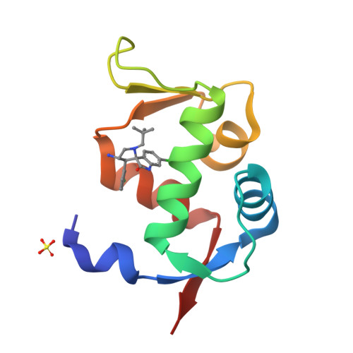
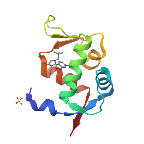
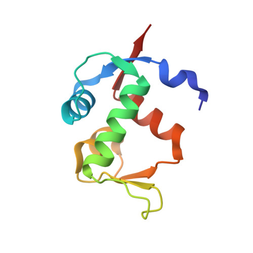
Novel Spiro[3H-indole-3,2 -pyrrolidin]-2(1H)-one Inhibitors of the MDM2-p53 Interaction: HDM2 (MDM2) in complex with compound 6b


Experimental Data Snapshot

  • Method: X-RAY DIFFRACTION
  • Resolution: 1.73 Å
  • R-Value Free: 0.273 
  • R-Value Work: 0.241 
  • R-Value Observed: 0.244 

wwPDB Validation   3D Report Full Report


Ligand Structure Quality Assessment 


This is version 1.3 of the entry. See complete history


Literature

Discovery of Novel Spiro[3H-indole-3,2'-pyrrolidin]-2(1H)-one Compounds as Chemically Stable and Orally Active Inhibitors of the MDM2-p53 Interaction.

Gollner, A.Rudolph, D.Arnhof, H.Bauer, M.Blake, S.M.Boehmelt, G.Cockroft, X.L.Dahmann, G.Ettmayer, P.Gerstberger, T.Karolyi-Oezguer, J.Kessler, D.Kofink, C.Ramharter, J.Rinnenthal, J.Savchenko, A.Schnitzer, R.Weinstabl, H.Weyer-Czernilofsky, U.Wunberg, T.McConnell, D.B.

(2016) J Med Chem 59: 10147-10162

  • DOI: https://doi.org/10.1021/acs.jmedchem.6b00900
  • Primary Citation of Related Structures:  
    5LAV, 5LAW, 5LAY, 5LAZ

  • PubMed Abstract: 

    Scaffold modification based on Wang's pioneering MDM2-p53 inhibitors led to novel, chemically stable spiro-oxindole compounds bearing a spiro[3H-indole-3,2'-pyrrolidin]-2(1H)-one scaffold that are not prone to epimerization as observed for the initial spiro[3H-indole-3,3'-pyrrolidin]-2(1H)-one scaffold. Further structure-based optimization inspired by natural product architectures led to a complex fused ring system ideally suited to bind to the MDM2 protein and to interrupt its protein-protein interaction (PPI) with TP53. The compounds are highly selective and show in vivo efficacy in a SJSA-1 xenograft model even when given as a single dose as demonstrated for 4-[(3S,3'S,3'aS,5'R,6'aS)-6-chloro-3'-(3-chloro-2-fluorophenyl)-1'-(cyclopropylmethyl)-2-oxo-1,2,3',3'a,4',5',6',6'a-octahydro-1'H-spiro[indole-3,2'-pyrrolo[3,2-b]pyrrole]-5'-yl]benzoic acid (BI-0252).


  • Organizational Affiliation

    Boehringer Ingelheim RCV GmbH & Co. KG , Dr. Boehringer-Gasse 5-11, A-1121 Vienna, Austria.


Macromolecules
Find similar proteins by:  (by identity cutoff)  |  3D Structure
Entity ID: 1
MoleculeChains Sequence LengthOrganismDetailsImage
E3 ubiquitin-protein ligase Mdm293Homo sapiensMutation(s): 0 
Gene Names: MDM2
EC: 6.3.2 (PDB Primary Data), 2.3.2.27 (UniProt)
UniProt & NIH Common Fund Data Resources
Find proteins for Q00987 (Homo sapiens)
Explore Q00987 
Go to UniProtKB:  Q00987
PHAROS:  Q00987
GTEx:  ENSG00000135679 
Entity Groups  
Sequence Clusters30% Identity50% Identity70% Identity90% Identity95% Identity100% Identity
UniProt GroupQ00987
Sequence Annotations
Expand
  • Reference Sequence
Small Molecules
Ligands 2 Unique
IDChains Name / Formula / InChI Key2D Diagram3D Interactions
6SK
Query on 6SK

Download Ideal Coordinates CCD File 
B [auth A](3~{S},3'~{S},4'~{S})-4'-azanyl-6-chloranyl-3'-(3-chlorophenyl)-1'-(2,2-dimethylpropyl)spiro[1~{H}-indole-3,2'-pyrrolidine]-2-one
C22 H25 Cl2 N3 O
CWDGHXCEKKZGEC-ZWCUEREDSA-N
SO4
Query on SO4

Download Ideal Coordinates CCD File 
C [auth A]SULFATE ION
O4 S
QAOWNCQODCNURD-UHFFFAOYSA-L
Experimental Data & Validation

Experimental Data

  • Method: X-RAY DIFFRACTION
  • Resolution: 1.73 Å
  • R-Value Free: 0.273 
  • R-Value Work: 0.241 
  • R-Value Observed: 0.244 
  • Space Group: P 61 2 2
Unit Cell:
Length ( Å )Angle ( ˚ )
a = 56.651α = 90
b = 56.651β = 90
c = 104.085γ = 120
Software Package:
Software NamePurpose
REFMACrefinement
PDB_EXTRACTdata extraction
XDSdata reduction
XSCALEdata scaling
REFMACphasing

Structure Validation

View Full Validation Report


Ligand Structure Quality Assessment 


Entry History 

Deposition Data

Revision History  (Full details and data files)

  • Version 1.0: 2016-11-02
    Type: Initial release
  • Version 1.1: 2016-12-21
    Changes: Database references
  • Version 1.2: 2018-04-25
    Changes: Advisory, Data collection, Structure summary
  • Version 1.3: 2024-05-08
    Changes: Advisory, Data collection, Database references