5L8K | pdb_00005l8k

Aurora-A kinase domain in complex with vNAR-D01 (crystal form 2)


Experimental Data Snapshot

  • Method: X-RAY DIFFRACTION
  • Resolution: 1.79 Å
  • R-Value Free: 
    0.235 (Depositor), 0.209 (DCC) 
  • R-Value Work: 
    0.190 (Depositor) 
  • R-Value Observed: 
    0.192 (Depositor) 

Starting Models: experimental
View more details

wwPDB Validation 3D Report Full Report

Validation slider image for 5L8K

Ligand Structure Quality Assessment 


This is version 2.1 of the entry. See complete history

Literature

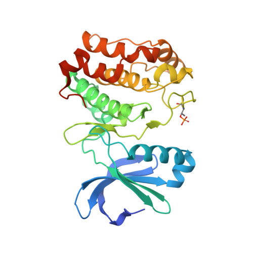
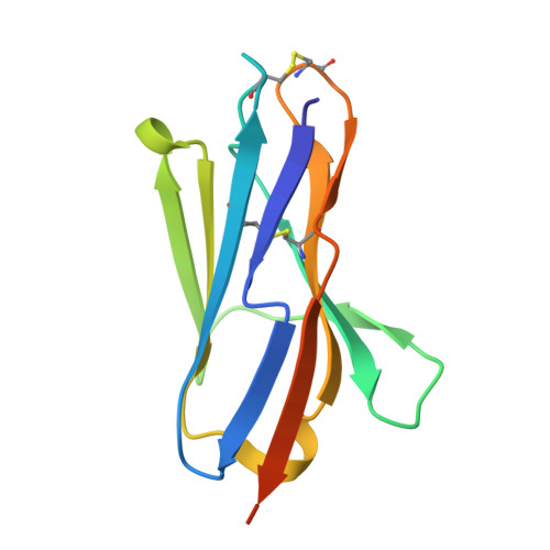
Allosteric inhibition of Aurora-A kinase by a synthetic vNAR domain.

Burgess, S.G.Oleksy, A.Cavazza, T.Richards, M.W.Vernos, I.Matthews, D.Bayliss, R.

(2016) Open Biol 6

  • DOI: https://doi.org/10.1098/rsob.160089
  • Primary Citation Related Structures: 
    5L8J, 5L8K, 5L8L

  • PubMed Abstract: 

    The vast majority of clinically approved protein kinase inhibitors target the ATP-binding pocket directly. Consequently, many inhibitors have broad selectivity profiles and most have significant off-target effects. Allosteric inhibitors are generally more selective, but are difficult to identify because allosteric binding sites are often unknown or poorly characterized. Aurora-A is activated through binding of TPX2 to an allosteric site on the kinase catalytic domain, and this knowledge could be exploited to generate an inhibitor. Here, we generated an allosteric inhibitor of Aurora-A kinase based on a synthetic, vNAR single domain scaffold, vNAR-D01. Biochemical studies and a crystal structure of the Aurora-A/vNAR-D01 complex show that the vNAR domain overlaps with the TPX2 binding site. In contrast with the binding of TPX2, which stabilizes an active conformation of the kinase, binding of the vNAR domain stabilizes an inactive conformation, in which the αC-helix is distorted, the canonical Lys-Glu salt bridge is broken and the regulatory (R-) spine is disrupted by an additional hydrophobic side chain from the activation loop. These studies illustrate how single domain antibodies can be used to characterize the regulatory mechanisms of kinases and provide a rational basis for structure-guided design of allosteric Aurora-A kinase inhibitors.


  • Organizational Affiliation
    • Astbury Centre for Structural Molecular Biology, Faculty of Biological Sciences, University of Leeds, Leeds LS2 9JT, UK Department of Molecular and Cell Biology, University of Leicester, Leicester LE1 9HN, UK.

Macromolecule Content 

  • Total Structure Weight: 46.97 kDa 
  • Atom Count: 3,246 
  • Modeled Residue Count: 372 
  • Deposited Residue Count: 402 
  • Unique protein chains: 2

Macromolecules

Find similar proteins by:|  3D Structure
Entity ID: 1
MoleculeChains  Sequence LengthOrganismDetailsImage
Aurora kinase A285Homo sapiensMutation(s): 2 
Gene Names: AURKAAIKAIRK1ARK1AURAAYK1BTAKIAK1STK15STK6
EC: 2.7.11.1
UniProt & NIH Common Fund Data Resources
Find proteins for O14965 (Homo sapiens)
Explore O14965 
Go to UniProtKB:  O14965
PHAROS:  O14965
GTEx:  ENSG00000087586 
Entity Groups
Sequence Clusters30% Identity50% Identity70% Identity90% Identity95% Identity100% Identity
UniProt GroupO14965
Sequence Annotations
Expand
Reference Sequence
Find similar proteins by:|  3D Structure
Entity ID: 2
MoleculeChains  Sequence LengthOrganismDetailsImage
New antigen receptor variable domain117Orectolobus maculatusMutation(s): 1 
UniProt
Find proteins for Q8JJ25 (Orectolobus maculatus)
Explore Q8JJ25 
Go to UniProtKB:  Q8JJ25
Entity Groups
Sequence Clusters30% Identity50% Identity70% Identity90% Identity95% Identity100% Identity
UniProt GroupQ8JJ25
Sequence Annotations
Expand
Reference Sequence

Small Molecules

Ligands 3 Unique
IDChains Name / Formula / InChI Key2D Diagram3D Interactions
ADP

Query on ADP


Download:Ideal Coordinates CCD File
C [auth A]ADENOSINE-5'-DIPHOSPHATE
C10 H15 N5 O10 P2
XTWYTFMLZFPYCI-KQYNXXCUSA-N
SO4

Query on SO4


Download:Ideal Coordinates CCD File
D [auth A],
O [auth B]
SULFATE ION
O4 S
QAOWNCQODCNURD-UHFFFAOYSA-L
EDO

Query on EDO


Download:Ideal Coordinates CCD File
E [auth A]
F [auth A]
G [auth A]
H [auth A]
I [auth A]
E [auth A],
F [auth A],
G [auth A],
H [auth A],
I [auth A],
J [auth A],
K [auth A],
L [auth A],
M [auth A],
N [auth A]
1,2-ETHANEDIOL
C2 H6 O2
LYCAIKOWRPUZTN-UHFFFAOYSA-N
Modified Residues  1 Unique
IDChains TypeFormula2D DiagramParent
TPO
Query on TPO
A
L-PEPTIDE LINKINGC4 H10 N O6 PTHR

Experimental Data & Validation

Experimental Data

  • Method: X-RAY DIFFRACTION
  • Resolution: 1.79 Å
  • R-Value Free:  0.235 (Depositor), 0.209 (DCC) 
  • R-Value Work:  0.190 (Depositor) 
  • R-Value Observed: 0.192 (Depositor) 
Space Group: P 21 21 2
Unit Cell:
Length ( Å )Angle ( ˚ )
a = 88.7α = 90
b = 109.72β = 90
c = 45.76γ = 90
Software Package:
Software NamePurpose
PHENIXrefinement
xia2data reduction
xia2data scaling
PHENIXphasing

Structure Validation

View Full Validation Report


Ligand Structure Quality Assessment 


Entry History 

& Funding Information

Deposition Data


Funding OrganizationLocationGrant Number
Cancer Research UKUnited KingdomC24461/A13231

Revision History  (Full details and data files)

  • Version 1.0: 2016-07-20
    Type: Initial release
  • Version 1.1: 2016-07-27
    Changes: Database references
  • Version 2.0: 2024-01-10
    Changes: Atomic model, Data collection, Database references, Refinement description
  • Version 2.1: 2024-10-23
    Changes: Structure summary