5L7U | pdb_00005l7u

Crystal structure of BvGH123 with bound GalNAc


Experimental Data Snapshot

  • Method: X-RAY DIFFRACTION
  • Resolution: 2.10 Å
  • R-Value Free: 
    0.227 (Depositor), 0.205 (DCC) 
  • R-Value Work: 
    0.193 (Depositor) 
  • R-Value Observed: 
    0.194 (Depositor) 

Starting Model: experimental
View more details

wwPDB Validation 3D Report Full Report

Validation slider image for 5L7U

Ligand Structure Quality Assessment 


This is version 1.3 of the entry. See complete history

Literature

Structural and mechanistic insights into a Bacteroides vulgatus retaining N-acetyl-beta-galactosaminidase that uses neighbouring group participation.

Roth, C.Petricevic, M.John, A.Goddard-Borger, E.D.Davies, G.J.Williams, S.J.

(2016) Chem Commun (Camb) 52: 11096-11099

  • DOI: https://doi.org/10.1039/c6cc04649e
  • Primary Citation Related Structures: 
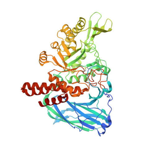
    5L7R, 5L7U, 5L7V

  • PubMed Abstract: 

    Bacteroides vulgatus is a member of the human microbiota whose abundance is increased in patients with Crohn's disease. We show that a B. vulgatus glycoside hydrolase from the carbohydrate active enzyme family GH123, BvGH123, is an N-acetyl-β-galactosaminidase that acts with retention of stereochemistry, and, through a 3-D structure in complex with Gal-thiazoline, provide evidence in support of a neighbouring group participation mechanism.


  • Organizational Affiliation
    • Department of Chemistry, University of York, Heslington, York, UK. gideon.davies@york.ac.uk.

Macromolecule Content 

  • Total Structure Weight: 133.13 kDa 
  • Atom Count: 9,250 
  • Modeled Residue Count: 1,115 
  • Deposited Residue Count: 1,156 
  • Unique protein chains: 1

Macromolecules

Find similar proteins by:|  3D Structure
Entity ID: 1
MoleculeChains  Sequence LengthOrganismDetailsImage
Glycoside hydrolase
A, B
578Phocaeicola vulgatus ATCC 8482Mutation(s): 0 
Gene Names: BVU_2198
UniProt
Find proteins for A6L2E5 (Phocaeicola vulgatus (strain ATCC 8482 / DSM 1447 / JCM 5826 / CCUG 4940 / NBRC 14291 / NCTC 11154))
Explore A6L2E5 
Go to UniProtKB:  A6L2E5
Entity Groups
Sequence Clusters30% Identity50% Identity70% Identity90% Identity95% Identity100% Identity
UniProt GroupA6L2E5
Sequence Annotations
Expand
Reference Sequence

Experimental Data & Validation

Experimental Data

  • Method: X-RAY DIFFRACTION
  • Resolution: 2.10 Å
  • R-Value Free:  0.227 (Depositor), 0.205 (DCC) 
  • R-Value Work:  0.193 (Depositor) 
  • R-Value Observed: 0.194 (Depositor) 
Space Group: P 21 21 21
Unit Cell:
Length ( Å )Angle ( ˚ )
a = 55.345α = 90
b = 142.207β = 90
c = 145.806γ = 90
Software Package:
Software NamePurpose
REFMACrefinement
PDB_EXTRACTdata extraction
XDSdata reduction
XDSdata scaling
MOLREPphasing

Structure Validation

View Full Validation Report


Ligand Structure Quality Assessment 


Entry History 

Deposition Data

Revision History  (Full details and data files)

  • Version 1.0: 2017-03-22
    Type: Initial release
  • Version 1.1: 2020-07-29
    Type: Remediation
    Reason: Carbohydrate remediation
    Changes: Data collection, Derived calculations, Structure summary
  • Version 1.2: 2024-01-10
    Changes: Data collection, Database references, Refinement description, Structure summary
  • Version 1.3: 2024-10-23
    Changes: Structure summary