5L7J | pdb_00005l7j

Crystal Structure of Elp3 from Dehalococcoides mccartyi


Experimental Data Snapshot

  • Method: X-RAY DIFFRACTION
  • Resolution: 2.15 Å
  • R-Value Free: 
    0.219 (Depositor), 0.230 (DCC) 
  • R-Value Work: 
    0.191 (Depositor), 0.206 (DCC) 
  • R-Value Observed: 
    0.193 (Depositor) 

wwPDB Validation 3D Report Full Report

Validation slider image for 5L7J

This is version 2.0 of the entry. See complete history

Literature

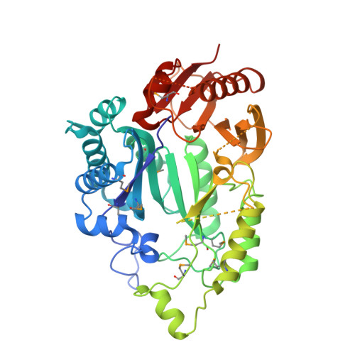
Structural basis for tRNA modification by Elp3 from Dehalococcoides mccartyi.

Glatt, S.Zabel, R.Kolaj-Robin, O.Onuma, O.F.Baudin, F.Graziadei, A.Taverniti, V.Lin, T.Y.Baymann, F.Seraphin, B.Breunig, K.D.Muller, C.W.

(2016) Nat Struct Mol Biol 23: 794-802

  • DOI: https://doi.org/10.1038/nsmb.3265
  • Primary Citation Related Structures: 
    5L7J, 5L7L

  • PubMed Abstract: 

    During translation elongation, decoding is based on the recognition of codons by corresponding tRNA anticodon triplets. Molecular mechanisms that regulate global protein synthesis via specific base modifications in tRNA anticodons are receiving increasing attention. The conserved eukaryotic Elongator complex specifically modifies uridines located in the wobble base position of tRNAs. Mutations in Elongator subunits are associated with certain neurodegenerative diseases and cancer. Here we present the crystal structure of D. mccartyi Elp3 (DmcElp3) at 2.15-Å resolution. Our results reveal an unexpected arrangement of Elp3 lysine acetyltransferase (KAT) and radical S-adenosyl methionine (SAM) domains, which share a large interface and form a composite active site and tRNA-binding pocket, with an iron-sulfur cluster located in the dimerization interface of two DmcElp3 molecules. Structure-guided mutagenesis studies of yeast Elp3 confirmed the relevance of our findings for eukaryotic Elp3s and should aid in understanding the cellular functions and pathophysiological roles of Elongator.


  • Organizational Affiliation
    • European Molecular Biology Laboratory, Structural and Computational Biology Unit, Heidelberg, Germany.

Macromolecule Content 

  • Total Structure Weight: 52.01 kDa 
  • Atom Count: 3,216 
  • Modeled Residue Count: 406 
  • Deposited Residue Count: 459 
  • Unique protein chains: 1

Macromolecules

Find similar proteins by:|  3D Structure
Entity ID: 1
MoleculeChains  Sequence LengthOrganismDetailsImage
ELP3 family459Dehalococcoides mccartyi BTF08Mutation(s): 1 
Gene Names: btf_573
EC: 2.3.1.311
UniProt
Find proteins for A0A1C7D1B6 (Dehalococcoides mccartyi BTF08)
Explore A0A1C7D1B6 
Go to UniProtKB:  A0A1C7D1B6
Entity Groups
Sequence Clusters30% Identity50% Identity70% Identity90% Identity95% Identity100% Identity
UniProt GroupA0A1C7D1B6
Sequence Annotations
Expand
Reference Sequence

Small Molecules

Modified Residues  1 Unique
IDChains TypeFormula2D DiagramParent
MSE
Query on MSE
A
L-PEPTIDE LINKINGC5 H11 N O2 SeMET

Experimental Data & Validation

Experimental Data

  • Method: X-RAY DIFFRACTION
  • Resolution: 2.15 Å
  • R-Value Free:  0.219 (Depositor), 0.230 (DCC) 
  • R-Value Work:  0.191 (Depositor), 0.206 (DCC) 
  • R-Value Observed: 0.193 (Depositor) 
Space Group: C 2 2 21
Unit Cell:
Length ( Å )Angle ( ˚ )
a = 75.46α = 90
b = 158.58β = 90
c = 93.58γ = 90
Software Package:
Software NamePurpose
PHENIXrefinement
XDSdata reduction
XSCALEdata scaling
autoSHARPphasing

Structure Validation

View Full Validation Report


Entry History 

Deposition Data

Revision History  (Full details and data files)

  • Version 1.0: 2016-08-03
    Type: Initial release
  • Version 1.1: 2016-08-10
    Changes: Database references
  • Version 1.2: 2016-09-21
    Changes: Database references
  • Version 2.0: 2024-11-13
    Changes: Atomic model, Data collection, Database references, Derived calculations, Structure summary