5L7E

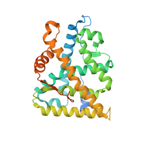
MCR IN COMPLEX WITH ligand


Experimental Data Snapshot

  • Method: X-RAY DIFFRACTION
  • Resolution: 1.86 Å
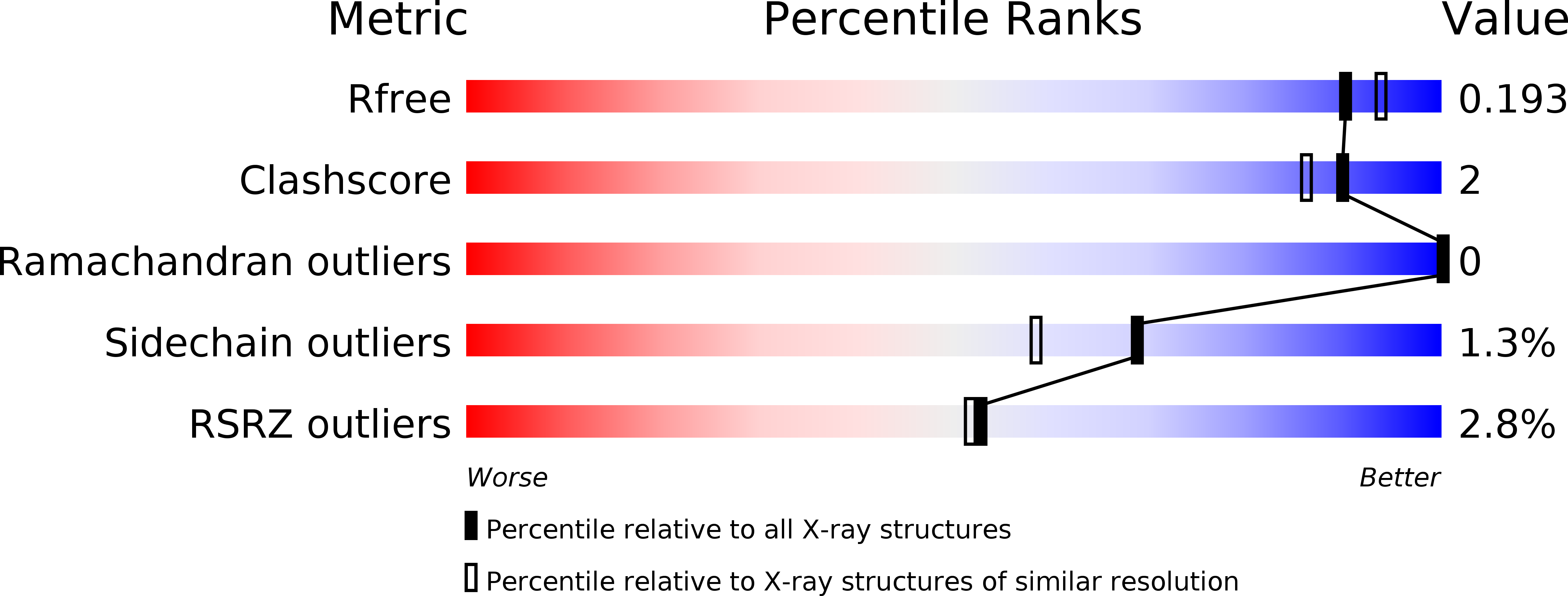
  • R-Value Free: 0.197 
  • R-Value Work: 0.180 
  • R-Value Observed: 0.181 

wwPDB Validation   3D Report Full Report


Ligand Structure Quality Assessment 


This is version 1.3 of the entry. See complete history


Literature

Structure-Based Drug Design of Mineralocorticoid Receptor Antagonists to Explore Oxosteroid Receptor Selectivity.

Nordqvist, A.O'Mahony, G.Friden-Saxin, M.Fredenwall, M.Hogner, A.Granberg, K.L.Aagaard, A.Backstrom, S.Gunnarsson, A.Kaminski, T.Xue, Y.Dellsen, A.Hansson, E.Hansson, P.Ivarsson, I.Karlsson, U.Bamberg, K.Hermansson, M.Georgsson, J.Lindmark, B.Edman, K.

(2017) ChemMedChem 12: 50-65

  • DOI: https://doi.org/10.1002/cmdc.201600529
  • Primary Citation of Related Structures:  
    5L7E, 5L7G, 5L7H

  • PubMed Abstract: 

    The mineralocorticoid receptor (MR) is a nuclear hormone receptor involved in the regulation of body fluid and electrolyte homeostasis. In this study we explore selectivity triggers for a series of nonsteroidal MR antagonists to improve selectivity over other members of the oxosteroid receptor family. A biaryl sulfonamide compound was identified in a high-throughput screening (HTS) campaign. The compound bound to MR with pK i =6.6, but displayed poor selectivity over the glucocorticoid receptor (GR) and the progesterone receptor (PR). Following X-ray crystallography of MR in complex with the HTS hit, a compound library was designed that explored an induced-fit hypothesis that required movement of the Met852 side chain. An improvement in MR selectivity of 11- to 79-fold over PR and 23- to 234-fold over GR was obtained. Given the U-shaped binding conformation, macrocyclizations were explored, yielding a macrocycle that bound to MR with pK i =7.3. Two protein-ligand X-ray structures were determined, confirming the hypothesized binding mode for the designed compounds.


  • Organizational Affiliation

    Cardiovascular and Metabolic Diseases, Innovative Medicines and Early Development Biotech Unit, AstraZeneca, Pepparedsleden 1, Mölndal, 43183, Sweden.


Macromolecules
Find similar proteins by:  (by identity cutoff)  |  3D Structure
Entity ID: 1
MoleculeChains Sequence LengthOrganismDetailsImage
Mineralocorticoid receptor305Homo sapiensMutation(s): 0 
Gene Names: NR3C2MCRMLR
UniProt & NIH Common Fund Data Resources
Find proteins for P08235 (Homo sapiens)
Explore P08235 
Go to UniProtKB:  P08235
PHAROS:  P08235
GTEx:  ENSG00000151623 
Entity Groups  
Sequence Clusters30% Identity50% Identity70% Identity90% Identity95% Identity100% Identity
UniProt GroupP08235
Sequence Annotations
Expand
  • Reference Sequence

Find similar proteins by:  Sequence   |   3D Structure  

Entity ID: 2
MoleculeChains Sequence LengthOrganismDetailsImage
NCOA1 peptide10Homo sapiensMutation(s): 0 
EC: 2.3.1.48
UniProt & NIH Common Fund Data Resources
Find proteins for Q15788 (Homo sapiens)
Explore Q15788 
Go to UniProtKB:  Q15788
PHAROS:  Q15788
GTEx:  ENSG00000084676 
Entity Groups  
Sequence Clusters30% Identity50% Identity70% Identity90% Identity95% Identity100% Identity
UniProt GroupQ15788
Sequence Annotations
Expand
  • Reference Sequence
Small Molecules
Ligands 5 Unique
IDChains Name / Formula / InChI Key2D Diagram3D Interactions
6Q0
Query on 6Q0

Download Ideal Coordinates CCD File 
H [auth A]~{N}-[[4-(3,5-dimethyl-1,2-oxazol-4-yl)phenyl]methyl]benzenesulfonamide
C18 H18 N2 O3 S
RZFYAEOEPOUGRI-UHFFFAOYSA-N
NHE
Query on NHE

Download Ideal Coordinates CCD File 
C [auth A]2-[N-CYCLOHEXYLAMINO]ETHANE SULFONIC ACID
C8 H17 N O3 S
MKWKNSIESPFAQN-UHFFFAOYSA-N
MPD
Query on MPD

Download Ideal Coordinates CCD File 
D [auth A](4S)-2-METHYL-2,4-PENTANEDIOL
C6 H14 O2
SVTBMSDMJJWYQN-YFKPBYRVSA-N
SO4
Query on SO4

Download Ideal Coordinates CCD File 
E [auth A]SULFATE ION
O4 S
QAOWNCQODCNURD-UHFFFAOYSA-L
EDO
Query on EDO

Download Ideal Coordinates CCD File 
F [auth A],
G [auth A]
1,2-ETHANEDIOL
C2 H6 O2
LYCAIKOWRPUZTN-UHFFFAOYSA-N
Experimental Data & Validation

Experimental Data

  • Method: X-RAY DIFFRACTION
  • Resolution: 1.86 Å
  • R-Value Free: 0.197 
  • R-Value Work: 0.180 
  • R-Value Observed: 0.181 
  • Space Group: P 21 21 21
Unit Cell:
Length ( Å )Angle ( ˚ )
a = 48.62α = 90
b = 77.83β = 90
c = 78.65γ = 90
Software Package:
Software NamePurpose
BUSTERrefinement
XDSdata reduction
SCALAdata scaling

Structure Validation

View Full Validation Report


Ligand Structure Quality Assessment 


Entry History 

Deposition Data

Revision History  (Full details and data files)

  • Version 1.0: 2016-12-07
    Type: Initial release
  • Version 1.1: 2017-01-18
    Changes: Database references
  • Version 1.2: 2019-10-16
    Changes: Data collection
  • Version 1.3: 2024-05-08
    Changes: Data collection, Database references