5L4L | pdb_00005l4l

polyketide ketoreductase SimC7 - ternary complex with NADP+ and 7-oxo-SD8


Experimental Data Snapshot

  • Method: X-RAY DIFFRACTION
  • Resolution: 1.20 Å
  • R-Value Free: 
    0.149 (Depositor), 0.149 (DCC) 
  • R-Value Work: 
    0.126 (Depositor), 0.125 (DCC) 
  • R-Value Observed: 
    0.127 (Depositor) 

Starting Model: experimental
View more details

wwPDB Validation 3D Report Full Report

Validation slider image for 5L4L

Ligand Structure Quality Assessment 


This is version 2.1 of the entry. See complete history

Literature

Substrate-Assisted Catalysis in Polyketide Reduction Proceeds via a Phenolate Intermediate.

Schafer, M.Stevenson, C.E.Wilkinson, B.Lawson, D.M.Buttner, M.J.

(2016) Cell Chem Biol 23: 1091-1097

  • DOI: https://doi.org/10.1016/j.chembiol.2016.07.018
  • Primary Citation Related Structures: 
    5L3Z, 5L40, 5L45, 5L4L

  • PubMed Abstract: 

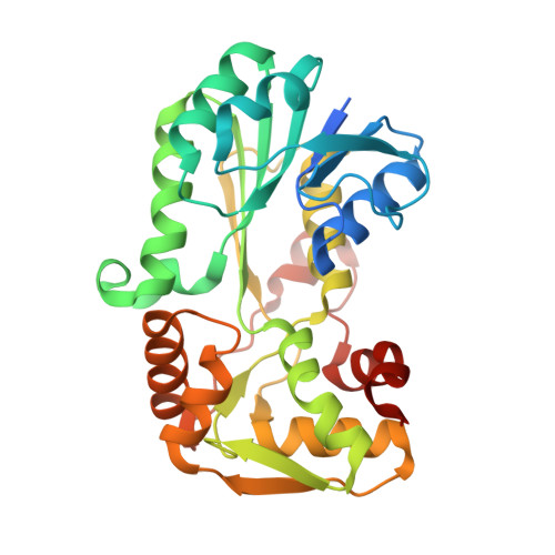
    SimC7 is a polyketide ketoreductase involved in biosynthesis of the angucyclinone moiety of the gyrase inhibitor simocyclinone D8 (SD8). SimC7, which belongs to the short-chain dehydrogenase/reductase (SDR) superfamily, catalyzes reduction of the C-7 carbonyl of the angucyclinone, and the resulting hydroxyl is essential for antibiotic activity. SimC7 shares little sequence similarity with characterized ketoreductases, suggesting it might have a distinct mechanism. To investigate this possibility, we determined the structures of SimC7 alone, with NADP(+), and with NADP(+) and the substrate 7-oxo-SD8. These structures show that SimC7 is distinct from previously characterized polyketide ketoreductases, lacking the conserved catalytic triad, including the active-site tyrosine that acts as central acid-base catalyst in canonical SDR proteins. Taken together with functional analyses of active-site mutants, our data suggest that SimC7 catalyzes a substrate-assisted, two-step reaction for reduction of the C-7 carbonyl group involving intramolecular transfer of a substrate-derived proton to generate a phenolate intermediate.


  • Organizational Affiliation
    • Department of Molecular Microbiology, John Innes Centre, Norwich Research Park, Norwich NR4 7UH, UK.

Macromolecule Content 

  • Total Structure Weight: 34.04 kDa 
  • Atom Count: 2,689 
  • Modeled Residue Count: 278 
  • Deposited Residue Count: 304 
  • Unique protein chains: 1

Macromolecules

Find similar proteins by:|  3D Structure
Entity ID: 1
MoleculeChains  Sequence LengthOrganismDetailsImage
SimC7304Streptomyces antibioticusMutation(s): 0 
Gene Names: simC7
UniProt
Find proteins for G9VYV4 (Streptomyces antibioticus)
Explore G9VYV4 
Go to UniProtKB:  G9VYV4
Entity Groups
Sequence Clusters30% Identity50% Identity70% Identity90% Identity95% Identity100% Identity
UniProt GroupG9VYV4
Sequence Annotations
Expand
Reference Sequence

Experimental Data & Validation

Experimental Data

  • Method: X-RAY DIFFRACTION
  • Resolution: 1.20 Å
  • R-Value Free:  0.149 (Depositor), 0.149 (DCC) 
  • R-Value Work:  0.126 (Depositor), 0.125 (DCC) 
  • R-Value Observed: 0.127 (Depositor) 
Space Group: P 21 21 21
Unit Cell:
Length ( Å )Angle ( ˚ )
a = 51.93α = 90
b = 53.66β = 90
c = 102.27γ = 90
Software Package:
Software NamePurpose
Aimlessdata scaling
REFMACrefinement
PDB_EXTRACTdata extraction
PHASERphasing
XDSdata reduction

Structure Validation

View Full Validation Report


Ligand Structure Quality Assessment 


Entry History 

& Funding Information

Deposition Data


Funding OrganizationLocationGrant Number
Biotechnology and Biological Sciences Research CouncilUnited KingdomBB/I002197/1
Biotechnology and Biological Sciences Research CouncilUnited KingdomBB/J004561/1
Biotechnology and Biological Sciences Research CouncilUnited KingdomDoctoral Training Partnership

Revision History  (Full details and data files)

  • Version 1.0: 2016-10-05
    Type: Initial release
  • Version 2.0: 2017-08-30
    Changes: Atomic model, Author supporting evidence, Derived calculations
  • Version 2.1: 2024-01-10
    Changes: Data collection, Database references, Refinement description