5L2J | pdb_00005l2j

Crystal Structure of human CD1b in complex with C36-GMM


Experimental Data Snapshot

  • Method: X-RAY DIFFRACTION
  • Resolution: 1.65 Å
  • R-Value Free: 
    0.199 (Depositor), 0.201 (DCC) 
  • R-Value Work: 
    0.172 (Depositor) 
  • R-Value Observed: 
    0.174 (Depositor) 

Starting Model: experimental
View more details

wwPDB Validation 3D Report Full Report

Validation slider image for 5L2J

Ligand Structure Quality Assessment 


This is version 2.2 of the entry. See complete history

Literature

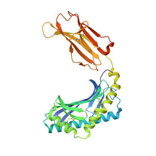
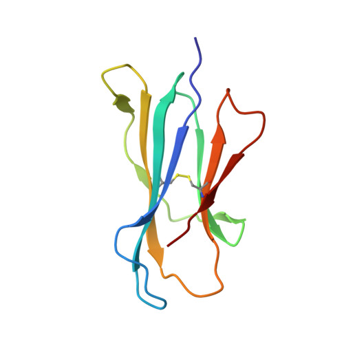
T cell receptor recognition of CD1b presenting a mycobacterial glycolipid.

Gras, S.Van Rhijn, I.Shahine, A.Cheng, T.Y.Bhati, M.Tan, L.L.Halim, H.Tuttle, K.D.Gapin, L.Le Nours, J.Moody, D.B.Rossjohn, J.

(2016) Nat Commun 7: 13257-13257

  • DOI: https://doi.org/10.1038/ncomms13257
  • Primary Citation Related Structures: 
    5L2J, 5L2K

  • PubMed Abstract: 

    CD1 proteins present microbial lipids to T cells. Germline-encoded mycolyl lipid-reactive (GEM) T cells with conserved αβ T cell receptors (TCRs) recognize CD1b presenting mycobacterial mycolates. As the molecular basis underpinning TCR recognition of CD1b remains unknown, here we determine the structure of a GEM TCR bound to CD1b presenting glucose-6-O-monomycolate (GMM). The GEM TCR docks centrally above CD1b, whereby the conserved TCR α-chain extensively contacts CD1b and GMM. Through mutagenesis and study of T cells from tuberculosis patients, we identify a consensus CD1b footprint of TCRs present among GEM T cells. Using both the TCR α- and β-chains as tweezers to surround and grip the glucose moiety of GMM, GEM TCRs create a highly specific mechanism for recognizing this mycobacterial glycolipid.


  • Organizational Affiliation
    • Infection and Immunity Program, Biomedicine Discovery Institute, Monash University, Clayton, Victoria, Australia.

Macromolecule Content 

  • Total Structure Weight: 48.09 kDa 
  • Atom Count: 3,618 
  • Modeled Residue Count: 377 
  • Deposited Residue Count: 398 
  • Unique protein chains: 2

Macromolecules

Find similar proteins by:|  3D Structure
Entity ID: 1
MoleculeChains  Sequence LengthOrganismDetailsImage
T-cell surface glycoprotein CD1b300Homo sapiensMutation(s): 0 
Gene Names: CD1B
UniProt & NIH Common Fund Data Resources
Find proteins for P29016 (Homo sapiens)
Explore P29016 
Go to UniProtKB:  P29016
PHAROS:  P29016
GTEx:  ENSG00000158485 
Entity Groups
Sequence Clusters30% Identity50% Identity70% Identity90% Identity95% Identity100% Identity
UniProt GroupP29016
Glycosylation
Glycosylation Sites: 2Go to GlyGen: P29016-1
Sequence Annotations
Expand
Reference Sequence
Find similar proteins by:|  3D Structure
Entity ID: 2
MoleculeChains  Sequence LengthOrganismDetailsImage
Beta-2-microglobulin98Homo sapiensMutation(s): 0 
Gene Names: B2MCDABP0092HDCMA22P
UniProt & NIH Common Fund Data Resources
Find proteins for P61769 (Homo sapiens)
Explore P61769 
Go to UniProtKB:  P61769
PHAROS:  P61769
GTEx:  ENSG00000166710 
Entity Groups
Sequence Clusters30% Identity50% Identity70% Identity90% Identity95% Identity100% Identity
UniProt GroupP61769
Sequence Annotations
Expand
Reference Sequence

Oligosaccharides

Help  
Entity ID: 3
MoleculeChains Length2D Diagram GlycosylationD Interactions
alpha-D-mannopyranose-(1-3)-[alpha-D-mannopyranose-(1-6)]beta-D-mannopyranose-(1-4)-2-acetamido-2-deoxy-beta-D-glucopyranose-(1-4)-[alpha-L-fucopyranose-(1-3)][alpha-L-fucopyranose-(1-6)]2-acetamido-2-deoxy-beta-D-glucopyranose
C
7N-Glycosylation
Glycosylation Resources
GlyTouCan: G11460AB
GlyCosmos: G11460AB
GlyGen: G11460AB

Small Molecules

Ligands 6 Unique
IDChains Name / Formula / InChI Key2D Diagram3D Interactions
70E

Query on 70E


Download:Ideal Coordinates CCD File
J [auth A]6-O-[(2R,3R)-3-hydroxy-2-tetradecyldocosanoyl]-alpha-L-idopyranose
C42 H82 O8
YOCZDGRJDQLKHW-GWLFWXKWSA-N
6UL

Query on 6UL


Download:Ideal Coordinates CCD File
D [auth A]TETRACOSYL PALMITATE
C40 H80 O2
KKTAIMZGSAQXRE-UHFFFAOYSA-N
NAG

Query on NAG


Download:Ideal Coordinates CCD File
E [auth A]2-acetamido-2-deoxy-beta-D-glucopyranose
C8 H15 N O6
OVRNDRQMDRJTHS-FMDGEEDCSA-N
ACT

Query on ACT


Download:Ideal Coordinates CCD File
F [auth A],
G [auth A]
ACETATE ION
C2 H3 O2
QTBSBXVTEAMEQO-UHFFFAOYSA-M
CL

Query on CL


Download:Ideal Coordinates CCD File
H [auth A],
I [auth A]
CHLORIDE ION
Cl
VEXZGXHMUGYJMC-UHFFFAOYSA-M
NA

Query on NA


Download:Ideal Coordinates CCD File
K [auth B]SODIUM ION
Na
FKNQFGJONOIPTF-UHFFFAOYSA-N

Experimental Data & Validation

Experimental Data

  • Method: X-RAY DIFFRACTION
  • Resolution: 1.65 Å
  • R-Value Free:  0.199 (Depositor), 0.201 (DCC) 
  • R-Value Work:  0.172 (Depositor) 
  • R-Value Observed: 0.174 (Depositor) 
Space Group: P 21 21 21
Unit Cell:
Length ( Å )Angle ( ˚ )
a = 57.66α = 90
b = 78.11β = 90
c = 91.33γ = 90
Software Package:
Software NamePurpose
BUSTERrefinement
Aimlessdata scaling
PHASERphasing
PDB_EXTRACTdata extraction
XDSdata reduction

Structure Validation

View Full Validation Report


Ligand Structure Quality Assessment 


Entry History 

Deposition Data

Revision History  (Full details and data files)

  • Version 1.0: 2016-11-16
    Type: Initial release
  • Version 1.1: 2016-12-07
    Changes: Other
  • Version 2.0: 2020-07-29
    Type: Remediation
    Reason: Carbohydrate remediation
    Changes: Advisory, Atomic model, Data collection, Derived calculations, Structure summary
  • Version 2.1: 2023-10-04
    Changes: Data collection, Database references, Derived calculations, Refinement description, Structure summary
  • Version 2.2: 2024-11-20
    Changes: Structure summary