5L22 | pdb_00005l22

PrtD T1SS ABC transporter


Experimental Data Snapshot

  • Method: X-RAY DIFFRACTION
  • Resolution: 3.15 Å
  • R-Value Free: 
    0.276 (Depositor), 0.280 (DCC) 
  • R-Value Work: 
    0.223 (Depositor), 0.230 (DCC) 
  • R-Value Observed: 
    0.226 (Depositor) 

wwPDB Validation   3D Report Full Report


Ligand Structure Quality Assessment 

Created with Raphaël 2.3.0Worse 01 BetterLigand structure goodness of fit to experimental dataBest fitted ADPClick on this verticalbar to view details

This is version 1.2 of the entry. See complete history


Literature

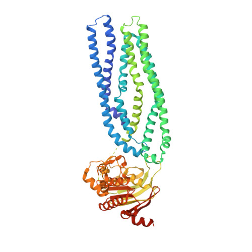
Structure of a Type-1 Secretion System ABC Transporter.

Morgan, J.L.Acheson, J.F.Zimmer, J.

(2017) Structure 25: 522-529

  • DOI: https://doi.org/10.1016/j.str.2017.01.010
  • Primary Citation of Related Structures:  
    5L22

  • PubMed Abstract: 

    Type-1 secretion systems (T1SSs) represent a widespread mode of protein secretion across the cell envelope in Gram-negative bacteria. The T1SS is composed of an inner-membrane ABC transporter, a periplasmic membrane-fusion protein, and an outer-membrane porin. These three components assemble into a complex spanning both membranes and providing a conduit for the translocation of unfolded polypeptides. We show that ATP hydrolysis and assembly of the entire T1SS complex is necessary for protein secretion. Furthermore, we present a 3.15-Å crystal structure of AaPrtD, the ABC transporter found in the Aquifex aeolicus T1SS. The structure suggests a substrate entry window just above the transporter's nucleotide binding domains. In addition, highly kinked transmembrane helices, which frame a narrow channel not observed in canonical peptide transporters, are likely involved in substrate translocation. Overall, the AaPrtD structure supports a polypeptide transport mechanism distinct from alternating access.


  • Organizational Affiliation

    Department of Molecular Physiology and Biological Physics, University of Virginia School of Medicine, 480 Ray C. Hunt Drive, Charlottesville, VA 22908, USA. Electronic address: jlm2qp@virginia.edu.


Macromolecules
Find similar proteins by:  (by identity cutoff)  |  3D Structure
Entity ID: 1
MoleculeChains Sequence LengthOrganismDetailsImage
ABC transporter (HlyB subfamily)A [auth B],
B [auth A]
572Aquifex aeolicus VF5Mutation(s): 0 
Gene Names: abcT5aq_1097
Membrane Entity: Yes 
UniProt
Find proteins for O67184 (Aquifex aeolicus (strain VF5))
Explore O67184 
Go to UniProtKB:  O67184
Entity Groups  
Sequence Clusters30% Identity50% Identity70% Identity90% Identity95% Identity100% Identity
UniProt GroupO67184
Sequence Annotations
Expand
  • Reference Sequence
Experimental Data & Validation

Experimental Data

  • Method: X-RAY DIFFRACTION
  • Resolution: 3.15 Å
  • R-Value Free:  0.276 (Depositor), 0.280 (DCC) 
  • R-Value Work:  0.223 (Depositor), 0.230 (DCC) 
  • R-Value Observed: 0.226 (Depositor) 
Space Group: C 1 2 1
Unit Cell:
Length ( Å )Angle ( ˚ )
a = 118.672α = 90
b = 97.938β = 100.47
c = 179.823γ = 90
Software Package:
Software NamePurpose
PHENIXrefinement
XDSdata reduction
Aimlessdata scaling
SHELXCDphasing

Structure Validation

View Full Validation Report


Ligand Structure Quality Assessment 

Created with Raphaël 2.3.0Worse 01 BetterLigand structure goodness of fit to experimental dataBest fitted ADPClick on this verticalbar to view details

Entry History 

Deposition Data

Revision History  (Full details and data files)

  • Version 1.0: 2017-03-08
    Type: Initial release
  • Version 1.1: 2017-03-22
    Changes: Database references
  • Version 1.2: 2024-03-06
    Changes: Data collection, Database references, Derived calculations