5KZV | pdb_00005kzv

Crystal structure of the xenopus Smoothened cysteine-rich domain (CRD) in complex with 20(S)-hydroxycholesterol


Experimental Data Snapshot

  • Method: X-RAY DIFFRACTION
  • Resolution: 1.62 Å
  • R-Value Free: 
    0.207 (Depositor), 0.211 (DCC) 
  • R-Value Work: 
    0.174 (Depositor), 0.177 (DCC) 
  • R-Value Observed: 
    0.176 (Depositor) 

Starting Model: experimental
View more details

wwPDB Validation 3D Report Full Report

Validation slider image for 5KZV

Ligand Structure Quality Assessment 


This is version 1.5 of the entry. See complete history

Literature

Cellular Cholesterol Directly Activates Smoothened in Hedgehog Signaling.

Huang, P.Nedelcu, D.Watanabe, M.Jao, C.Kim, Y.Liu, J.Salic, A.

(2016) Cell 166: 1176-1187.e14

  • DOI: https://doi.org/10.1016/j.cell.2016.08.003
  • Primary Citation Related Structures: 
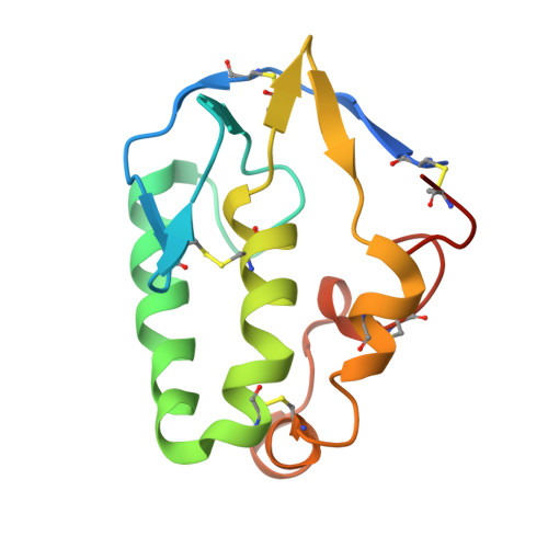
    5KZV, 5KZY, 5KZZ

  • PubMed Abstract: 

    In vertebrates, sterols are necessary for Hedgehog signaling, a pathway critical in embryogenesis and cancer. Sterols activate the membrane protein Smoothened by binding its extracellular, cysteine-rich domain (CRD). Major unanswered questions concern the nature of the endogenous, activating sterol and the mechanism by which it regulates Smoothened. We report crystal structures of CRD complexed with sterols and alone, revealing that sterols induce a dramatic conformational change of the binding site, which is sufficient for Smoothened activation and is unique among CRD-containing receptors. We demonstrate that Hedgehog signaling requires sterol binding to Smoothened and define key residues for sterol recognition and activity. We also show that cholesterol itself binds and activates Smoothened. Furthermore, the effect of oxysterols is abolished in Smoothened mutants that retain activation by cholesterol and Hedgehog. We propose that the endogenous Smoothened activator is cholesterol, not oxysterols, and that vertebrate Hedgehog signaling controls Smoothened by regulating its access to cholesterol.


  • Organizational Affiliation
    • Department of Cell Biology, Harvard Medical School, 240 Longwood Avenue, Boston, MA 02115, USA.

Macromolecule Content 

  • Total Structure Weight: 14.39 kDa 
  • Atom Count: 1,167 
  • Modeled Residue Count: 117 
  • Deposited Residue Count: 127 
  • Unique protein chains: 1

Macromolecules

Find similar proteins by:|  3D Structure
Entity ID: 1
MoleculeChains  Sequence LengthOrganismDetailsImage
Smoothened127Xenopus laevisMutation(s): 0 
Gene Names: Smo
UniProt
Find proteins for Q98SW5 (Xenopus laevis)
Explore Q98SW5 
Go to UniProtKB:  Q98SW5
Entity Groups
Sequence Clusters30% Identity50% Identity70% Identity90% Identity95% Identity100% Identity
UniProt GroupQ98SW5
Sequence Annotations
Expand
Reference Sequence

Small Molecules

Ligands 1 Unique
IDChains Name / Formula / InChI Key2D Diagram3D Interactions
HCD

Query on HCD


Download:Ideal Coordinates CCD File
B [auth A](3alpha,8alpha)-cholest-5-ene-3,20-diol
C27 H46 O2
MCKLJFJEQRYRQT-APGJSSKUSA-N

Experimental Data & Validation

Experimental Data

  • Method: X-RAY DIFFRACTION
  • Resolution: 1.62 Å
  • R-Value Free:  0.207 (Depositor), 0.211 (DCC) 
  • R-Value Work:  0.174 (Depositor), 0.177 (DCC) 
  • R-Value Observed: 0.176 (Depositor) 
Space Group: C 1 2 1
Unit Cell:
Length ( Å )Angle ( ˚ )
a = 73.248α = 90
b = 28.349β = 104.27
c = 61.929γ = 90
Software Package:
Software NamePurpose
PHENIXrefinement
HKL-2000data scaling
PDB_EXTRACTdata extraction
HKL-2000data reduction
PHASERphasing

Structure Validation

View Full Validation Report


Ligand Structure Quality Assessment 


Entry History 

& Funding Information

Deposition Data


Funding OrganizationLocationGrant Number
National Institutes of Health/National Institute of General Medical Sciences (NIH/NIGMS)United StatesRO1GM092924
National Institutes of Health/National Institute of General Medical Sciences (NIH/NIGMS)United StatesRO1GM110041

Revision History  (Full details and data files)

  • Version 1.0: 2016-08-17
    Type: Initial release
  • Version 1.1: 2016-09-07
    Changes: Database references
  • Version 1.2: 2017-09-06
    Changes: Author supporting evidence, Derived calculations
  • Version 1.3: 2019-12-25
    Changes: Author supporting evidence
  • Version 1.4: 2023-10-04
    Changes: Data collection, Database references, Refinement description
  • Version 1.5: 2024-11-20
    Changes: Structure summary