5KZM | pdb_00005kzm

Crystal structure of Tryptophan synthase alpha-beta chain complex from Francisella tularensis


Experimental Data Snapshot

  • Method: X-RAY DIFFRACTION
  • Resolution: 2.80 Å
  • R-Value Free: 
    0.235 (Depositor), 0.240 (DCC) 
  • R-Value Work: 
    0.183 (Depositor), 0.182 (DCC) 
  • R-Value Observed: 
    0.186 (Depositor) 

wwPDB Validation 3D Report Full Report

Validation slider image for 5KZM

This is version 2.2 of the entry. See complete history

Literature

Conservation of the structure and function of bacterial tryptophan synthases.

Michalska, K.Gale, J.Joachimiak, G.Chang, C.Hatzos-Skintges, C.Nocek, B.Johnston, S.E.Bigelow, L.Bajrami, B.Jedrzejczak, R.P.Wellington, S.Hung, D.T.Nag, P.P.Fisher, S.L.Endres, M.Joachimiak, A.

(2019) IUCrJ 6: 649-664

  • DOI: https://doi.org/10.1107/S2052252519005955
  • Primary Citation Related Structures: 
    5KIN, 5KZM

  • PubMed Abstract: 

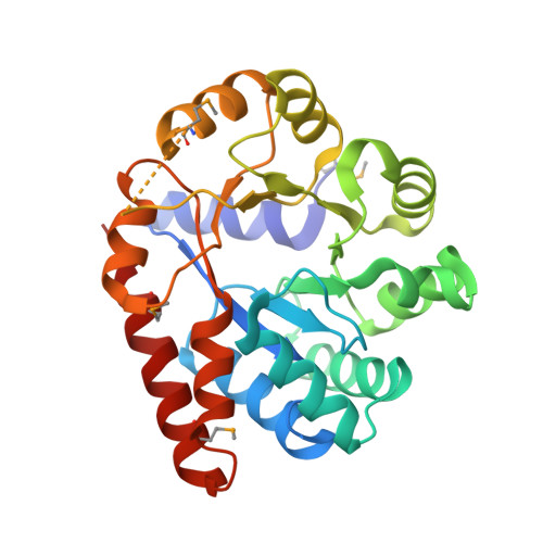
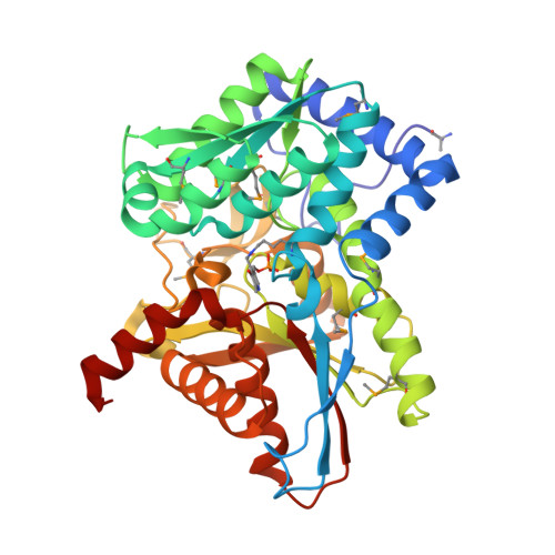
    Tryptophan biosynthesis is one of the most characterized processes in bacteria, in which the enzymes from Salmonella typhimurium and Escherichia coli serve as model systems. Tryptophan synthase (TrpAB) catalyzes the final two steps of tryptophan biosynthesis in plants, fungi and bacteria. This pyridoxal 5'-phosphate (PLP)-dependent enzyme consists of two protein chains, α (TrpA) and β (TrpB), functioning as a linear αββα heterotetrameric complex containing two TrpAB units. The reaction has a complicated, multistep mechanism resulting in the β-replacement of the hydroxyl group of l-serine with an indole moiety. Recent studies have shown that functional TrpAB is required for the survival of pathogenic bacteria in macrophages and for evading host defense. Therefore, TrpAB is a promising target for drug discovery, as its orthologs include enzymes from the important human pathogens Streptococcus pneumoniae , Legionella pneumophila and Francisella tularensis , the causative agents of pneumonia, legionnaires' disease and tularemia, respectively. However, specific biochemical and structural properties of the TrpABs from these organisms have not been investigated. To fill the important phylogenetic gaps in the understanding of TrpABs and to uncover unique features of TrpAB orthologs to spearhead future drug-discovery efforts, the TrpABs from L. pneumophila , F. tularensis and S. pneumoniae have been characterized. In addition to kinetic properties and inhibitor-sensitivity data, structural information gathered using X-ray crystallo-graphy is presented. The enzymes show remarkable structural conservation, but at the same time display local differences in both their catalytic and allosteric sites that may be responsible for the observed differences in catalysis and inhibitor binding. This functional dissimilarity may be exploited in the design of species-specific enzyme inhibitors.


  • Organizational Affiliation
    • Center for Structural Genomics of Infectious Diseases, University of Chicago, Chicago, IL 60367, USA.

Macromolecule Content 

  • Total Structure Weight: 73.53 kDa 
  • Atom Count: 4,987 
  • Modeled Residue Count: 655 
  • Deposited Residue Count: 668 
  • Unique protein chains: 2

Macromolecules

Find similar proteins by:|  3D Structure
Entity ID: 1
MoleculeChains  Sequence LengthOrganismDetailsImage
Tryptophan synthase alpha chain272Francisella tularensis subsp. tularensis SCHU S4Mutation(s): 0 
Gene Names: trpAFTT_1772c
EC: 4.2.1.20
UniProt
Find proteins for Q5NE80 (Francisella tularensis subsp. tularensis (strain SCHU S4 / Schu 4))
Explore Q5NE80 
Go to UniProtKB:  Q5NE80
Entity Groups
Sequence Clusters30% Identity50% Identity70% Identity90% Identity95% Identity100% Identity
UniProt GroupQ5NE80
Sequence Annotations
Expand
Reference Sequence
Find similar proteins by:|  3D Structure
Entity ID: 2
MoleculeChains  Sequence LengthOrganismDetailsImage
Tryptophan synthase beta chain396Francisella tularensis subsp. tularensis SCHU S4Mutation(s): 0 
Gene Names: trpBFTT_1773c
EC: 4.2.1.20
UniProt
Find proteins for Q5NE79 (Francisella tularensis subsp. tularensis (strain SCHU S4 / Schu 4))
Explore Q5NE79 
Go to UniProtKB:  Q5NE79
Entity Groups
Sequence Clusters30% Identity50% Identity70% Identity90% Identity95% Identity100% Identity
UniProt GroupQ5NE79
Sequence Annotations
Expand
Reference Sequence

Small Molecules

Experimental Data & Validation

Experimental Data

  • Method: X-RAY DIFFRACTION
  • Resolution: 2.80 Å
  • R-Value Free:  0.235 (Depositor), 0.240 (DCC) 
  • R-Value Work:  0.183 (Depositor), 0.182 (DCC) 
  • R-Value Observed: 0.186 (Depositor) 
Space Group: C 2 2 21
Unit Cell:
Length ( Å )Angle ( ˚ )
a = 111.176α = 90
b = 171.986β = 90
c = 76.109γ = 90
Software Package:
Software NamePurpose
PHENIXrefinement
PDB_EXTRACTdata extraction
HKL-3000data reduction
HKL-3000data scaling
HKL-3000phasing

Structure Validation

View Full Validation Report


Entry History 

Revision History  (Full details and data files)

  • Version 1.0: 2016-08-10
    Type: Initial release
  • Version 1.1: 2016-09-07
    Changes: Database references, Structure summary
  • Version 2.0: 2018-05-09
    Changes: Advisory, Atomic model, Data collection, Database references, Derived calculations, Polymer sequence, Source and taxonomy, Structure summary
  • Version 2.1: 2019-09-18
    Changes: Data collection, Database references
  • Version 2.2: 2025-04-02
    Changes: Data collection, Database references, Derived calculations, Structure summary