5KZD | pdb_00005kzd

N-acetylneuraminate lyase from methicillin-resistant Staphylococcus aureus with bound sialic acid alditol


Experimental Data Snapshot

  • Method: X-RAY DIFFRACTION
  • Resolution: 2.33 Å
  • R-Value Free: 
    0.261 (Depositor), 0.262 (DCC) 
  • R-Value Work: 
    0.193 (Depositor), 0.198 (DCC) 
  • R-Value Observed: 
    0.196 (Depositor) 

wwPDB Validation 3D Report Full Report

Validation slider image for 5KZD

Ligand Structure Quality Assessment 


This is version 1.1 of the entry. See complete history

Literature

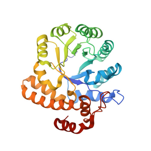
Structure and inhibition of N-acetylneuraminate lyase from methicillin-resistant Staphylococcus aureus.

North, R.A.Watson, A.J.Pearce, F.G.Muscroft-Taylor, A.C.Friemann, R.Fairbanks, A.J.Dobson, R.C.

(2016) FEBS Lett 590: 4414-4428

  • DOI: https://doi.org/10.1002/1873-3468.12462
  • Primary Citation Related Structures: 
    5KZD, 5KZE

  • PubMed Abstract: 

    N-Acetylneuraminate lyase is the first committed enzyme in the degradation of sialic acid by bacterial pathogens. In this study, we analyzed the kinetic parameters of N-acetylneuraminate lyase from methicillin-resistant Staphylococcus aureus (MRSA). We determined that the enzyme has a relatively high K M of 3.2 mm, suggesting that flux through the catabolic pathway is likely to be controlled by this enzyme. Our data indicate that sialic acid alditol, a known inhibitor of N-acetylneuraminate lyase enzymes, is a stronger inhibitor of MRSA N-acetylneuraminate lyase than of Clostridium perfringens N-acetylneuraminate lyase. Our analysis of the crystal structure of ligand-free and 2R-sialic acid alditol-bound MRSA N-acetylneuraminate lyase suggests that subtle dynamic differences in solution and/or altered binding interactions within the active site may account for species-specific inhibition.


  • Organizational Affiliation
    • Biomolecular Interaction Centre, School of Biological Sciences, University of Canterbury, Christchurch, New Zealand.

Macromolecule Content 

  • Total Structure Weight: 133.55 kDa 
  • Atom Count: 9,341 
  • Modeled Residue Count: 1,136 
  • Deposited Residue Count: 1,172 
  • Unique protein chains: 1

Macromolecules

Find similar proteins by:|  3D Structure
Entity ID: 1
MoleculeChains  Sequence LengthOrganismDetailsImage
N-acetylneuraminate lyase
A, B, C, D
293Staphylococcus aureus subsp. aureus USA300Mutation(s): 0 
Gene Names: nanASAUSA300_0315
EC: 4.1.3.3
UniProt
Find proteins for Q2G160 (Staphylococcus aureus (strain NCTC 8325 / PS 47))
Explore Q2G160 
Go to UniProtKB:  Q2G160
Entity Groups
Sequence Clusters30% Identity50% Identity70% Identity90% Identity95% Identity100% Identity
UniProt GroupQ2G160
Sequence Annotations
Expand
Reference Sequence

Experimental Data & Validation

Experimental Data

  • Method: X-RAY DIFFRACTION
  • Resolution: 2.33 Å
  • R-Value Free:  0.261 (Depositor), 0.262 (DCC) 
  • R-Value Work:  0.193 (Depositor), 0.198 (DCC) 
  • R-Value Observed: 0.196 (Depositor) 
Space Group: P 21 21 21
Unit Cell:
Length ( Å )Angle ( ˚ )
a = 81.477α = 90
b = 109.444β = 90
c = 130.752γ = 90
Software Package:
Software NamePurpose
Aimlessdata scaling
PHENIXrefinement
PDB_EXTRACTdata extraction
iMOSFLMdata reduction
PHASERphasing

Structure Validation

View Full Validation Report


Ligand Structure Quality Assessment 


Entry History 

Deposition Data

Revision History  (Full details and data files)

  • Version 1.0: 2017-01-11
    Type: Initial release
  • Version 1.1: 2024-03-06
    Changes: Data collection, Database references