5KUX | pdb_00005kux

Designed influenza hemagglutinin binding protein HSB.2


Experimental Data Snapshot

  • Method: X-RAY DIFFRACTION
  • Resolution: 1.80 Å
  • R-Value Free: 
    0.236 (Depositor), 0.238 (DCC) 
  • R-Value Work: 
    0.196 (Depositor), 0.199 (DCC) 
  • R-Value Observed: 
    0.198 (Depositor) 

Starting Model: experimental
View more details

wwPDB Validation 3D Report Full Report

Validation slider image for 5KUX

This is version 1.4 of the entry. See complete history

Literature

Computational design of trimeric influenza-neutralizing proteins targeting the hemagglutinin receptor binding site.

Strauch, E.M.Bernard, S.M.La, D.Bohn, A.J.Lee, P.S.Anderson, C.E.Nieusma, T.Holstein, C.A.Garcia, N.K.Hooper, K.A.Ravichandran, R.Nelson, J.W.Sheffler, W.Bloom, J.D.Lee, K.K.Ward, A.B.Yager, P.Fuller, D.H.Wilson, I.A.Baker, D.

(2017) Nat Biotechnol 35: 667-671

  • DOI: https://doi.org/10.1038/nbt.3907
  • Primary Citation Related Structures: 
    5KUX, 5KUY

  • PubMed Abstract: 

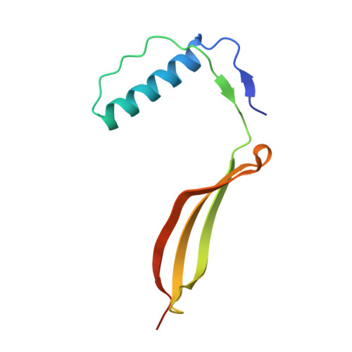
    Many viral surface glycoproteins and cell surface receptors are homo-oligomers, and thus can potentially be targeted by geometrically matched homo-oligomers that engage all subunits simultaneously to attain high avidity and/or lock subunits together. The adaptive immune system cannot generally employ this strategy since the individual antibody binding sites are not arranged with appropriate geometry to simultaneously engage multiple sites in a single target homo-oligomer. We describe a general strategy for the computational design of homo-oligomeric protein assemblies with binding functionality precisely matched to homo-oligomeric target sites. In the first step, a small protein is designed that binds a single site on the target. In the second step, the designed protein is assembled into a homo-oligomer such that the designed binding sites are aligned with the target sites. We use this approach to design high-avidity trimeric proteins that bind influenza A hemagglutinin (HA) at its conserved receptor binding site. The designed trimers can both capture and detect HA in a paper-based diagnostic format, neutralizes influenza in cell culture, and completely protects mice when given as a single dose 24 h before or after challenge with influenza.


  • Organizational Affiliation
    • Department of Biochemistry, University of Washington, Seattle, Washington, USA.

Macromolecule Content 

  • Total Structure Weight: 11.02 kDa 
  • Atom Count: 738 
  • Modeled Residue Count: 92 
  • Deposited Residue Count: 98 
  • Unique protein chains: 1

Macromolecules

Find similar proteins by:|  3D Structure
Entity ID: 1
MoleculeChains  Sequence LengthOrganismDetailsImage
Designed influenza inhibitor protein HSB.298synthetic constructMutation(s): 0 

Experimental Data & Validation

Experimental Data

  • Method: X-RAY DIFFRACTION
  • Resolution: 1.80 Å
  • R-Value Free:  0.236 (Depositor), 0.238 (DCC) 
  • R-Value Work:  0.196 (Depositor), 0.199 (DCC) 
  • R-Value Observed: 0.198 (Depositor) 
Space Group: P 32 2 1
Unit Cell:
Length ( Å )Angle ( ˚ )
a = 58.385α = 90
b = 58.385β = 90
c = 69.449γ = 120
Software Package:
Software NamePurpose
PHENIXrefinement
HKL-2000data reduction
SCALAdata scaling
PHASERphasing

Structure Validation

View Full Validation Report


Entry History 

& Funding Information

Deposition Data


Funding OrganizationLocationGrant Number
National Institutes of Health/National Institute Of Allergy and Infectious Diseases (NIH/NIAID)United StatesR56 AI117675

Revision History  (Full details and data files)

  • Version 1.0: 2017-06-14
    Type: Initial release
  • Version 1.1: 2017-06-28
    Changes: Database references
  • Version 1.2: 2017-07-26
    Changes: Author supporting evidence, Database references
  • Version 1.3: 2019-12-11
    Changes: Author supporting evidence
  • Version 1.4: 2023-10-04
    Changes: Data collection, Database references, Refinement description