5KTQ | pdb_00005ktq

LARGE FRAGMENT OF TAQ DNA POLYMERASE BOUND TO DCTP


Experimental Data Snapshot

  • Method: X-RAY DIFFRACTION
  • Resolution: 2.50 Å
  • R-Value Free: 
    0.280 (Depositor) 
  • R-Value Work: 
    0.217 (Depositor) 
  • R-Value Observed: 
    0.217 (Depositor) 

wwPDB Validation 3D Report Full Report

Validation slider image for 5KTQ

This is version 1.3 of the entry. See complete history

Literature

Crystal structures of the Klenow fragment of Thermus aquaticus DNA polymerase I complexed with deoxyribonucleoside triphosphates.

Li, Y.Kong, Y.Korolev, S.Waksman, G.

(1998) Protein Sci 7: 1116-1123

  • DOI: https://doi.org/10.1002/pro.5560070505
  • Primary Citation Related Structures: 
    5KTQ

  • PubMed Abstract: 

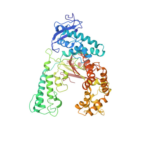
    The crystal structures of the Klenow fragment of the Thermus aquaticus DNA polymerase I (Klentaq1) complexed with four deoxyribonucleoside triphosphates (dNTP) have been determined to 2.5 A resolution. The dNTPs bind adjacent to the O helix of Klentaq1. The triphosphate moieties are at nearly identical positions in all four complexes and are anchored by three positively charged residues, Arg659, Lys663, and Arg587, and by two polar residues, His639 and Gln613. The configuration of the base moieties in the Klentaq1/dNTP complexes demonstrates variability suggesting that dNTP binding is primarily determined by recognition and binding of the phosphate moiety. However, when superimposed on the Taq polymerase/blunt end DNA complex structure (Eom et al., 1996), two of the dNTP/Klentaq1 structures demonstrate appropriate stacking of the nucleotide base with the 3' end of the DNA primer strand, suggesting that at least in these two binary complexes, the observed dNTP conformations are functionally relevant.


  • Organizational Affiliation
    • Department of Biochemistry and Molecular Biophysics, Washington University School of Medicine, Saint Louis, Missouri 63110, USA.

Macromolecule Content 

  • Total Structure Weight: 61.72 kDa 
  • Atom Count: 4,267 
  • Modeled Residue Count: 534 
  • Deposited Residue Count: 543 
  • Unique protein chains: 1

Macromolecules

Find similar proteins by:|  3D Structure
Entity ID: 1
MoleculeChains  Sequence LengthOrganismDetailsImage
PROTEIN (DNA POLYMERASE I)543Thermus aquaticusMutation(s): 0 
Gene Names: TAQ
EC: 2.7.7.7
UniProt
Find proteins for P19821 (Thermus aquaticus)
Explore P19821 
Go to UniProtKB:  P19821
Entity Groups
Sequence Clusters30% Identity50% Identity70% Identity90% Identity95% Identity100% Identity
UniProt GroupP19821
Sequence Annotations
Expand
Reference Sequence

Small Molecules

Ligands 1 Unique
IDChains Name / Formula / InChI Key2D Diagram3D Interactions
DCP

Query on DCP


Download:Ideal Coordinates CCD File
B [auth A]2'-DEOXYCYTIDINE-5'-TRIPHOSPHATE
C9 H16 N3 O13 P3
RGWHQCVHVJXOKC-SHYZEUOFSA-N

Experimental Data & Validation

Experimental Data

  • Method: X-RAY DIFFRACTION
  • Resolution: 2.50 Å
  • R-Value Free:  0.280 (Depositor) 
  • R-Value Work:  0.217 (Depositor) 
  • R-Value Observed: 0.217 (Depositor) 
Space Group: P 21 21 2
Unit Cell:
Length ( Å )Angle ( ˚ )
a = 109.4α = 90
b = 137β = 90
c = 45.8γ = 90
Software Package:
Software NamePurpose
X-PLORrefinement

Structure Validation

View Full Validation Report


Entry History 

Deposition Data

Revision History  (Full details and data files)

  • Version 1.0: 1998-09-30
    Type: Initial release
  • Version 1.1: 2008-04-27
    Changes: Version format compliance
  • Version 1.2: 2011-07-13
    Changes: Version format compliance
  • Version 1.3: 2024-03-06
    Changes: Data collection, Database references, Derived calculations