5KN0 | pdb_00005kn0

Native bovine skeletal calsequestrin, low-Ca2+ form

  • Classification: METAL BINDING PROTEIN
  • Organism(s): Bos taurus
  • Mutation(s): No 

  • Deposited: 2016-06-27 Released: 2016-10-05 
  • Deposition Author(s): Lewis, K.M., Byrd, S., Kang, C.
  • Funding Organization(s): National Institutes of Health/National Institute of General Medical Sciences (NIH/NIGMS), M.J. Murdock Charitable Trust

Experimental Data Snapshot

  • Method: X-RAY DIFFRACTION
  • Resolution: 2.73 Å
  • R-Value Free: 
    0.240 (Depositor), 0.241 (DCC) 
  • R-Value Work: 
    0.196 (Depositor), 0.199 (DCC) 
  • R-Value Observed: 
    0.198 (Depositor) 

Starting Model: experimental
View more details

wwPDB Validation 3D Report Full Report

Validation slider image for 5KN0

Ligand Structure Quality Assessment 


This is version 2.2 of the entry. See complete history

Literature

Characterization of Post-Translational Modifications to Calsequestrins of Cardiac and Skeletal Muscle.

Lewis, K.M.Munske, G.R.Byrd, S.S.Kang, J.Cho, H.J.Rios, E.Kang, C.

(2016) Int J Mol Sci 17

  • DOI: https://doi.org/10.3390/ijms17091539
  • Primary Citation Related Structures: 
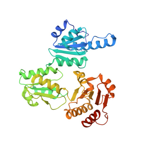
    5KN0, 5KN1, 5KN2, 5KN3

  • PubMed Abstract: 

    Calsequestrin is glycosylated and phosphorylated during its transit to its final destination in the junctional sarcoplasmic reticulum. To determine the significance and universal profile of these post-translational modifications to mammalian calsequestrin, we characterized, via mass spectrometry, the glycosylation and phosphorylation of skeletal muscle calsequestrin from cattle (B. taurus), lab mice (M. musculus) and lab rats (R. norvegicus) and cardiac muscle calsequestrin from cattle, lab rats and humans. On average, glycosylation of skeletal calsequestrin consisted of two N-acetylglucosamines and one mannose (GlcNAc₂Man₁), while cardiac calsequestrin had five additional mannoses (GlcNAc₂Man₆). Skeletal calsequestrin was not phosphorylated, while the C-terminal tails of cardiac calsequestrin contained between zero to two phosphoryls, indicating that phosphorylation of cardiac calsequestrin may be heterogeneous in vivo. Static light scattering experiments showed that the Ca(2+)-dependent polymerization capabilities of native bovine skeletal calsequestrin are enhanced, relative to the non-glycosylated, recombinant isoform, which our crystallographic studies suggest may be due to glycosylation providing a dynamic "guiderail"-like scaffold for calsequestrin polymerization. Glycosylation likely increases a polymerization/depolymerization response to changing Ca(2+) concentrations, and proper glycosylation, in turn, guarantees both effective Ca(2+) storage/buffering of the sarcoplasmic reticulum and localization of calsequestrin (Casq) at its target site.


  • Organizational Affiliation
    • Department of Chemistry, Washington State University, Pullman, WA 99164, USA. kevin_lewis@wsu.edu.

Macromolecule Content 

  • Total Structure Weight: 169.61 kDa 
  • Atom Count: 11,399 
  • Modeled Residue Count: 1,383 
  • Deposited Residue Count: 1,444 
  • Unique protein chains: 1

Macromolecules

Find similar proteins by:|  3D Structure
Entity ID: 1
MoleculeChains  Sequence LengthOrganismDetailsImage
Calsequestrin
A, B, C, D
361Bos taurusMutation(s): 0 
UniProt
Find proteins for Q05JF3 (Bos taurus)
Explore Q05JF3 
Go to UniProtKB:  Q05JF3
Entity Groups
Sequence Clusters30% Identity50% Identity70% Identity90% Identity95% Identity100% Identity
UniProt GroupQ05JF3
Glycosylation
Glycosylation Sites: 1
Sequence Annotations
Expand
Reference Sequence

Oligosaccharides

Help  
Entity ID: 2
MoleculeChains Length2D Diagram GlycosylationD Interactions
2-acetamido-2-deoxy-beta-D-glucopyranose-(1-4)-2-acetamido-2-deoxy-beta-D-glucopyranose
E
2N-Glycosylation
Glycosylation Resources
GlyTouCan: G42666HT
GlyCosmos: G42666HT
GlyGen: G42666HT
Entity ID: 3
MoleculeChains Length2D Diagram GlycosylationD Interactions
beta-D-mannopyranose-(1-4)-2-acetamido-2-deoxy-beta-D-glucopyranose-(1-4)-2-acetamido-2-deoxy-beta-D-glucopyranose
F
3N-Glycosylation
Glycosylation Resources
GlyTouCan: G15407YE
GlyCosmos: G15407YE
GlyGen: G15407YE

Small Molecules

Ligands 3 Unique
IDChains Name / Formula / InChI Key2D Diagram3D Interactions
NAG

Query on NAG


Download:Ideal Coordinates CCD File
G [auth A],
W [auth C]
2-acetamido-2-deoxy-beta-D-glucopyranose
C8 H15 N O6
OVRNDRQMDRJTHS-FMDGEEDCSA-N
MPD

Query on MPD


Download:Ideal Coordinates CCD File
CA [auth C]
DA [auth C]
EA [auth C]
JA [auth D]
KA [auth D]
CA [auth C],
DA [auth C],
EA [auth C],
JA [auth D],
KA [auth D],
M [auth A],
N [auth A],
T [auth B],
U [auth B],
V [auth B]
(4S)-2-METHYL-2,4-PENTANEDIOL
C6 H14 O2
SVTBMSDMJJWYQN-YFKPBYRVSA-N
CA

Query on CA


Download:Ideal Coordinates CCD File
AA [auth C]
BA [auth C]
FA [auth D]
GA [auth D]
H [auth A]
AA [auth C],
BA [auth C],
FA [auth D],
GA [auth D],
H [auth A],
HA [auth D],
I [auth A],
IA [auth D],
J [auth A],
K [auth A],
L [auth A],
O [auth B],
P [auth B],
Q [auth B],
R [auth B],
S [auth B],
X [auth C],
Y [auth C],
Z [auth C]
CALCIUM ION
Ca
BHPQYMZQTOCNFJ-UHFFFAOYSA-N

Experimental Data & Validation

Experimental Data

  • Method: X-RAY DIFFRACTION
  • Resolution: 2.73 Å
  • R-Value Free:  0.240 (Depositor), 0.241 (DCC) 
  • R-Value Work:  0.196 (Depositor), 0.199 (DCC) 
  • R-Value Observed: 0.198 (Depositor) 
Space Group: P 1
Unit Cell:
Length ( Å )Angle ( ˚ )
a = 60.342α = 71.12
b = 92.994β = 84.57
c = 101.849γ = 73.48
Software Package:
Software NamePurpose
PHENIXrefinement
HKL-2000data reduction
HKL-2000data scaling
PHENIXphasing

Structure Validation

View Full Validation Report


Ligand Structure Quality Assessment 


Entry History 

& Funding Information

Deposition Data


Funding OrganizationLocationGrant Number
National Institutes of Health/National Institute of General Medical Sciences (NIH/NIGMS)United States1R01GM11125401
M.J. Murdock Charitable TrustUnited States--

Revision History  (Full details and data files)

  • Version 1.0: 2016-10-05
    Type: Initial release
  • Version 1.1: 2017-09-27
    Changes: Author supporting evidence, Derived calculations
  • Version 1.2: 2019-12-25
    Changes: Author supporting evidence
  • Version 2.0: 2020-07-29
    Type: Remediation
    Reason: Carbohydrate remediation
    Changes: Atomic model, Data collection, Derived calculations, Structure summary
  • Version 2.1: 2023-09-27
    Changes: Data collection, Database references, Refinement description, Structure summary
  • Version 2.2: 2024-11-06
    Changes: Structure summary