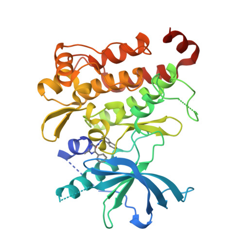
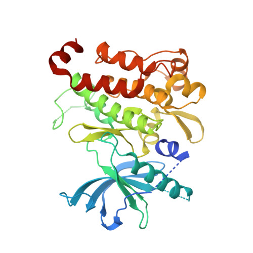
Structural characterization of nonactive site, TrkA-selective kinase inhibitors.
Su, H.P., Rickert, K., Burlein, C., Narayan, K., Bukhtiyarova, M., Hurzy, D.M., Stump, C.A., Zhang, X., Reid, J., Krasowska-Zoladek, A., Tummala, S., Shipman, J.M., Kornienko, M., Lemaire, P.A., Krosky, D., Heller, A., Achab, A., Chamberlin, C., Saradjian, P., Sauvagnat, B., Yang, X., Ziebell, M.R., Nickbarg, E., Sanders, J.M., Bilodeau, M.T., Carroll, S.S., Lumb, K.J., Soisson, S.M., Henze, D.A., Cooke, A.J.(2017) Proc Natl Acad Sci U S A 114: E297-E306
- PubMed: 28039433
- DOI: https://doi.org/10.1073/pnas.1611577114
- Primary Citation of Related Structures:
5KMI, 5KMJ, 5KMK, 5KML, 5KMM, 5KMN, 5KMO - PubMed Abstract:
Current therapies for chronic pain can have insufficient efficacy and lead to side effects, necessitating research of novel targets against pain. Although originally identified as an oncogene, Tropomyosin-related kinase A (TrkA) is linked to pain and elevated levels of NGF (the ligand for TrkA) are associated with chronic pain. Antibodies that block TrkA interaction with its ligand, NGF, are in clinical trials for pain relief. Here, we describe the identification of TrkA-specific inhibitors and the structural basis for their selectivity over other Trk family kinases. The X-ray structures reveal a binding site outside the kinase active site that uses residues from the kinase domain and the juxtamembrane region. Three modes of binding with the juxtamembrane region are characterized through a series of ligand-bound complexes. The structures indicate a critical pharmacophore on the compounds that leads to the distinct binding modes. The mode of interaction can allow TrkA selectivity over TrkB and TrkC or promiscuous, pan-Trk inhibition. This finding highlights the difficulty in characterizing the structure-activity relationship of a chemical series in the absence of structural information because of substantial differences in the interacting residues. These structures illustrate the flexibility of binding to sequences outside of-but adjacent to-the kinase domain of TrkA. This knowledge allows development of compounds with specificity for TrkA or the family of Trk proteins.
Organizational Affiliation:
Global Chemistry, Merck Research Laboratories (MRL), Merck & Co. Inc., West Point, PA 19486; hua-poo_su@merck.com.


