5KLH

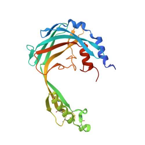
Crystal Structure of Trypanosoma brucei Procyclic Specific Surface Antigen-2


Experimental Data Snapshot

  • Method: X-RAY DIFFRACTION
  • Resolution: 1.65 Å
  • R-Value Free: 0.214 
  • R-Value Work: 0.177 
  • R-Value Observed: 0.179 

wwPDB Validation   3D Report Full Report


This is version 1.3 of the entry. See complete history


Literature

Structural characterization reveals a novel bilobed architecture for the ectodomains of insect stage expressed Trypanosoma brucei PSSA-2 and Trypanosoma congolense ISA.

Ramaswamy, R.Goomeshi Nobary, S.Eyford, B.A.Pearson, T.W.Boulanger, M.J.

(2016) Protein Sci 25: 2297-2302

  • DOI: https://doi.org/10.1002/pro.3053
  • Primary Citation of Related Structures:  
    5KLH, 5KMX

  • PubMed Abstract: 

    African trypanosomiasis, caused by parasites of the genus Trypanosoma, is a complex of devastating vector-borne diseases of humans and livestock in sub-Saharan Africa. Central to the pathogenesis of African trypanosomes is their transmission by the arthropod vector, Glossina spp. (tsetse fly). Intriguingly, the efficiency of parasite transmission through the vector is reduced following depletion of Trypanosoma brucei Procyclic-Specific Surface Antigen-2 (TbPSSA-2). To investigate the underlying molecular mechanism of TbPSSA-2, we determined the crystal structures of its ectodomain and that of its homolog T. congolense Insect Stage Antigen (TcISA) to resolutions of 1.65 Å and 2.45 Å, respectively using single wavelength anomalous dispersion. Both proteins adopt a novel bilobed architecture with the individual lobes displaying rotational flexibility around the central tether that suggest a potential mechanism for coordinating a binding partner. In support of this hypothesis, electron density consistent with a bound peptide was observed in the inter-lob cleft of a TcISA monomer. These first reported structures of insect stage transmembrane proteins expressed by African trypanosomes provide potentially valuable insight into the interface between parasite and tsetse vector.


  • Organizational Affiliation

    Department of Biochemistry and Microbiology, University of Victoria, Victoria, BC, Canada, V8W 3P6.


Macromolecules
Find similar proteins by:  (by identity cutoff)  |  3D Structure
Entity ID: 1
MoleculeChains Sequence LengthOrganismDetailsImage
Surface glycoprotein
A, B
231Trypanosoma bruceiMutation(s): 0 
UniProt
Find proteins for Q26806 (Trypanosoma brucei)
Explore Q26806 
Go to UniProtKB:  Q26806
Entity Groups  
Sequence Clusters30% Identity50% Identity70% Identity90% Identity95% Identity100% Identity
UniProt GroupQ26806
Sequence Annotations
Expand
  • Reference Sequence
Small Molecules
Ligands 1 Unique
IDChains Name / Formula / InChI Key2D Diagram3D Interactions
SO4
Query on SO4

Download Ideal Coordinates CCD File 
C [auth A]SULFATE ION
O4 S
QAOWNCQODCNURD-UHFFFAOYSA-L
Experimental Data & Validation

Experimental Data

  • Method: X-RAY DIFFRACTION
  • Resolution: 1.65 Å
  • R-Value Free: 0.214 
  • R-Value Work: 0.177 
  • R-Value Observed: 0.179 
  • Space Group: P 1
Unit Cell:
Length ( Å )Angle ( ˚ )
a = 31.82α = 73.55
b = 57.81β = 76.43
c = 61.24γ = 89.98
Software Package:
Software NamePurpose
PHENIXrefinement
PDB_EXTRACTdata extraction
iMOSFLMdata reduction
SCALAdata scaling
PHASERphasing

Structure Validation

View Full Validation Report


Entry History & Funding Information

Deposition Data


Funding OrganizationLocationGrant Number
Natural Sciences and Engineering Research Council (NSERC, Canada)Canada--

Revision History  (Full details and data files)

  • Version 1.0: 2017-07-05
    Type: Initial release
  • Version 1.1: 2017-07-26
    Changes: Author supporting evidence, Database references
  • Version 1.2: 2020-01-08
    Changes: Author supporting evidence
  • Version 1.3: 2024-11-06
    Changes: Data collection, Database references, Structure summary