5KKC | pdb_00005kkc

l-lactate dehydrogenase from rabbit muscle with the inhibitor 6DHNAD

  • Classification: OXIDOREDUCTASE
  • Organism(s): Oryctolagus cuniculus
  • Mutation(s): No 

  • Deposited: 2016-06-21 Released: 2016-11-02 
  • Deposition Author(s): Meneely, K.M., Moran, G.R., Lamb, A.L.
  • Funding Organization(s): National Science Foundation (NSF, United States), UWM Research Growth Initiative, National Institutes of Health/National Institute Of Allergy and Infectious Diseases (NIH/NIAID)

Experimental Data Snapshot

  • Method: X-RAY DIFFRACTION
  • Resolution: 1.86 Å
  • R-Value Free: 
    0.211 (Depositor), 0.214 (DCC) 
  • R-Value Work: 
    0.170 (Depositor), 0.172 (DCC) 
  • R-Value Observed: 
    0.170 (Depositor) 

Starting Model: experimental
View more details

wwPDB Validation 3D Report Full Report

Validation slider image for 5KKC

Ligand Structure Quality Assessment 


This is version 1.3 of the entry. See complete history

Literature

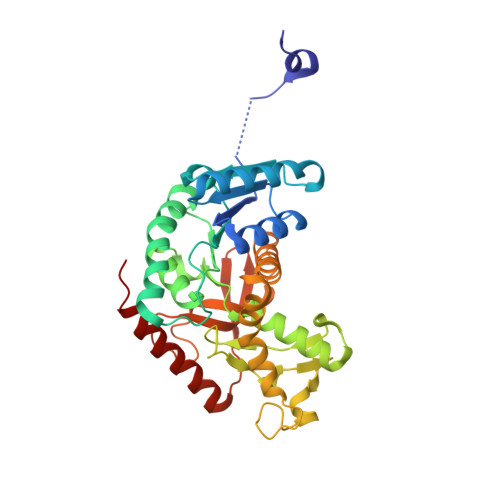
Ligand binding phenomena that pertain to the metabolic function of renalase.

Beaupre, B.A.Roman, J.V.Hoag, M.R.Meneely, K.M.Silvaggi, N.R.Lamb, A.L.Moran, G.R.

(2016) Arch Biochem Biophys 612: 46-56

  • DOI: https://doi.org/10.1016/j.abb.2016.10.011
  • Primary Citation Related Structures: 
    5KKA, 5KKC, 5KRQ

  • PubMed Abstract: 

    Renalase catalyzes the oxidation of isomers of β-NAD(P)H that carry the hydride in the 2 or 6 positions of the nicotinamide base to form β-NAD(P) + . This activity is thought to alleviate inhibition of multiple β-NAD(P)-dependent enzymes of primary and secondary metabolism by these isomers. Here we present evidence for a variety of ligand binding phenomena relevant to the function of renalase. We offer evidence of the potential for primary metabolism inhibition with structures of malate dehydrogenase and lactate dehydrogenase bound to the 6-dihydroNAD isomer. The previously observed preference of renalase from Pseudomonas for NAD-derived substrates over those derived from NADP is accounted for by the structure of the enzyme in complex with NADPH. We also show that nicotinamide nucleosides and mononucleotides reduced in the 2- and 6-positions are renalase substrates, but bind weakly. A seven-fold enhancement of acquisition (k red /K d ) for 6-dihydronicotinamide riboside was observed for human renalase in the presence of ADP. However, generally the addition of complement ligands, AMP for mononucleotide or ADP for nucleoside substrates, did not enhance the reductive half-reaction. Non-substrate nicotinamide nucleosides or nucleotides bind weakly suggesting that only β-NADH and β-NADPH compete with dinucleotide substrates for access to the active site.


  • Organizational Affiliation
    • Department of Chemistry and Biochemistry, University of Wisconsin-Milwaukee, 3210 N. Cramer St, Milwaukee, WI, 53211-3209, United States.

Macromolecule Content 

  • Total Structure Weight: 149.01 kDa 
  • Atom Count: 10,802 
  • Modeled Residue Count: 1,303 
  • Deposited Residue Count: 1,324 
  • Unique protein chains: 1

Macromolecules

Find similar proteins by:|  3D Structure
Entity ID: 1
MoleculeChains  Sequence LengthOrganismDetailsImage
L-lactate dehydrogenase A chain
A, B, C, D
331Oryctolagus cuniculusMutation(s): 0 
EC: 1.1.1.27
UniProt
Find proteins for P13491 (Oryctolagus cuniculus)
Explore P13491 
Go to UniProtKB:  P13491
Entity Groups
Sequence Clusters30% Identity50% Identity70% Identity90% Identity95% Identity100% Identity
UniProt GroupP13491
Sequence Annotations
Expand
Reference Sequence

Small Molecules

Ligands 2 Unique
IDChains Name / Formula / InChI Key2D Diagram3D Interactions
6V0

Query on 6V0


Download:Ideal Coordinates CCD File
E [auth A],
H [auth B],
J [auth C],
L [auth D]
[[(2~{R},3~{S},4~{R},5~{R})-5-(5-aminocarbonyl-2~{H}-pyridin-1-yl)-3,4-bis(oxidanyl)oxolan-2-yl]methoxy-oxidanyl-phosphoryl] [(2~{R},3~{S},4~{R},5~{R})-5-(6-aminopurin-9-yl)-3,4-bis(oxidanyl)oxolan-2-yl]methyl hydrogen phosphate
C21 H29 N7 O14 P2
QVQHBKNZCMZBKP-NNYOXOHSSA-N
SO4

Query on SO4


Download:Ideal Coordinates CCD File
F [auth A],
G [auth A],
I [auth B],
K [auth C],
M [auth D]
SULFATE ION
O4 S
QAOWNCQODCNURD-UHFFFAOYSA-L

Experimental Data & Validation

Experimental Data

  • Method: X-RAY DIFFRACTION
  • Resolution: 1.86 Å
  • R-Value Free:  0.211 (Depositor), 0.214 (DCC) 
  • R-Value Work:  0.170 (Depositor), 0.172 (DCC) 
  • R-Value Observed: 0.170 (Depositor) 
Space Group: P 1 21 1
Unit Cell:
Length ( Å )Angle ( ˚ )
a = 63.602α = 90
b = 126.085β = 99.94
c = 84.237γ = 90
Software Package:
Software NamePurpose
PHENIXrefinement
XDSdata reduction
XDSdata scaling
PHENIXphasing

Structure Validation

View Full Validation Report


Ligand Structure Quality Assessment 


Entry History 

& Funding Information

Deposition Data


Funding OrganizationLocationGrant Number
National Science Foundation (NSF, United States)United StatesCHE-1402475
UWM Research Growth InitiativeUnited States--
National Institutes of Health/National Institute Of Allergy and Infectious Diseases (NIH/NIAID)United StatesK02 AI093675
National Science Foundation (NSF, United States)United StatesCHE-1403293

Revision History  (Full details and data files)

  • Version 1.0: 2016-11-02
    Type: Initial release
  • Version 1.1: 2017-09-13
    Changes: Author supporting evidence, Database references, Derived calculations
  • Version 1.2: 2019-11-27
    Changes: Author supporting evidence
  • Version 1.3: 2023-09-27
    Changes: Data collection, Database references, Refinement description