5KHI | pdb_00005khi

HCN2 CNBD in complex with purine riboside-3', 5'-cyclic monophosphate (cPuMP)


Experimental Data Snapshot

  • Method: X-RAY DIFFRACTION
  • Resolution: 2.10 Å
  • R-Value Free: 
    0.249 (Depositor), 0.248 (DCC) 
  • R-Value Work: 
    0.201 (Depositor), 0.208 (DCC) 
  • R-Value Observed: 
    0.203 (Depositor) 

Starting Model: experimental
View more details

wwPDB Validation 3D Report Full Report

Validation slider image for 5KHI

Ligand Structure Quality Assessment 


This is version 1.2 of the entry. See complete history

Literature

Cyclic Purine and Pyrimidine Nucleotides Bind to the HCN2 Ion Channel and Variably Promote C-Terminal Domain Interactions and Opening.

Ng, L.C.Putrenko, I.Baronas, V.Van Petegem, F.Accili, E.A.

(2016) Structure 24: 1629-1642

  • DOI: https://doi.org/10.1016/j.str.2016.06.024
  • Primary Citation Related Structures: 
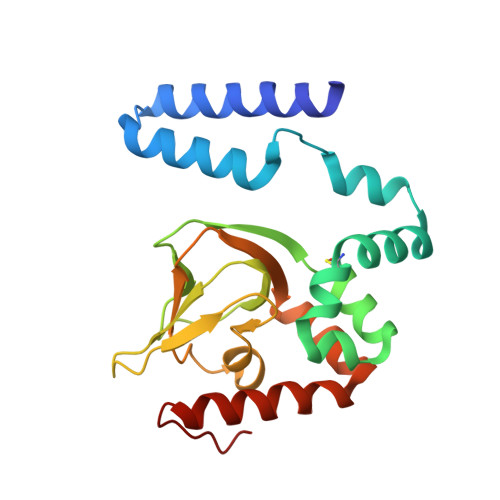
    5KHG, 5KHH, 5KHI, 5KHJ, 5KHK

  • PubMed Abstract: 

    Cyclic AMP is thought to facilitate the opening of the HCN2 channel by binding to a C-terminal domain and promoting or inhibiting interactions between subunits. Here, we correlated the ability of cyclic nucleotides to promote interactions of isolated HCN2 C-terminal domains in solution with their ability to facilitate channel opening. Cyclic IMP, a cyclic purine nucleotide, and cCMP, a cyclic pyrimidine nucleotide, bind to a C-terminal domain containing the cyclic nucleotide-binding domain but, in contrast to other cyclic nucleotides examined, fail to promote its oligomerization, and produce only modest facilitation of opening of the full-length channel. Comparisons between ligand bound structures identify a region between the sixth and seventh β strands and the distal C helix as important for facilitation and tight binding. We propose that promotion of interactions between the C-terminal domains by a given ligand contribute to its ability to facilitate opening of the full-length channel.


  • Organizational Affiliation
    • Department of Cellular and Physiological Sciences, University of British Columbia, Vancouver, BC V6T 1Z3, Canada.

Macromolecule Content 

  • Total Structure Weight: 24.2 kDa 
  • Atom Count: 1,683 
  • Modeled Residue Count: 201 
  • Deposited Residue Count: 204 
  • Unique protein chains: 1

Macromolecules

Find similar proteins by:|  3D Structure
Entity ID: 1
MoleculeChains  Sequence LengthOrganismDetailsImage
Potassium/sodium hyperpolarization-activated cyclic nucleotide-gated channel 2204Mus musculusMutation(s): 0 
Gene Names: Hcn2Bcng2Hac1
UniProt
Find proteins for O88703 (Mus musculus)
Explore O88703 
Go to UniProtKB:  O88703
Entity Groups
Sequence Clusters30% Identity50% Identity70% Identity90% Identity95% Identity100% Identity
UniProt GroupO88703
Sequence Annotations
Expand
Reference Sequence

Small Molecules

Ligands 1 Unique
IDChains Name / Formula / InChI Key2D Diagram3D Interactions
6SX

Query on 6SX


Download:Ideal Coordinates CCD File
B [auth A]Purine riboside-3',5'-cyclic monophosphate
C10 H11 N4 O6 P
AVSJXTVPIHQRPY-FDDDBJFASA-N
Modified Residues  1 Unique
IDChains TypeFormula2D DiagramParent
CSX
Query on CSX
A
L-PEPTIDE LINKINGC3 H7 N O3 SCYS

Experimental Data & Validation

Experimental Data

  • Method: X-RAY DIFFRACTION
  • Resolution: 2.10 Å
  • R-Value Free:  0.249 (Depositor), 0.248 (DCC) 
  • R-Value Work:  0.201 (Depositor), 0.208 (DCC) 
  • R-Value Observed: 0.203 (Depositor) 
Space Group: P 4 21 2
Unit Cell:
Length ( Å )Angle ( ˚ )
a = 96.278α = 90
b = 96.278β = 90
c = 46.763γ = 90
Software Package:
Software NamePurpose
XSCALEdata scaling
REFMACrefinement
PDB_EXTRACTdata extraction
XDSdata reduction
PHASERphasing

Structure Validation

View Full Validation Report


Ligand Structure Quality Assessment 


Entry History 

Deposition Data

Revision History  (Full details and data files)

  • Version 1.0: 2016-09-14
    Type: Initial release
  • Version 1.1: 2016-10-19
    Changes: Database references
  • Version 1.2: 2023-09-27
    Changes: Data collection, Database references, Derived calculations, Refinement description