5KBX | pdb_00005kbx

Co-crystal structure of the Saccharomyces cerevisiae histidine phosphotransfer signaling protein Ypd1 and the receiver domain of its downstream response regulator Ssk1


Experimental Data Snapshot

  • Method: X-RAY DIFFRACTION
  • Resolution: 2.80 Å
  • R-Value Free: 
    0.279 (Depositor), 0.275 (DCC) 
  • R-Value Work: 
    0.228 (Depositor), 0.229 (DCC) 
  • R-Value Observed: 
    0.233 (Depositor) 

wwPDB Validation 3D Report Full Report

Validation slider image for 5KBX

This is version 2.2 of the entry. See complete history

Literature

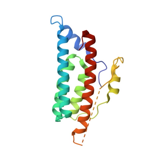
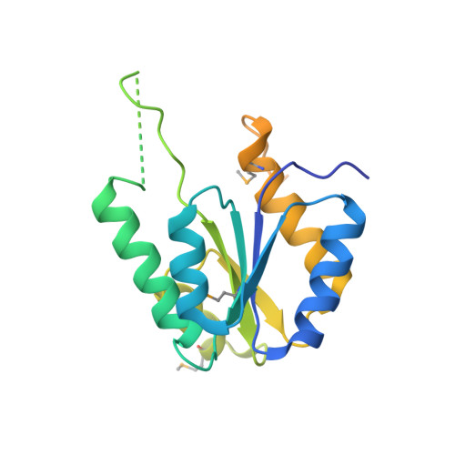
Insights revealed by the co-crystal structure of the Saccharomyces cerevisiae histidine phosphotransfer signaling protein Ypd1 and the receiver domain of its downstream response regulator Ssk1

Menon, S.K.Branscum, K.M.Foster, C.A.West, A.H.

To be published.

Macromolecule Content 

  • Total Structure Weight: 44.11 kDa 
  • Atom Count: 2,435 
  • Modeled Residue Count: 297 
  • Deposited Residue Count: 385 
  • Unique protein chains: 2

Macromolecules

Find similar proteins by:|  3D Structure
Entity ID: 1
MoleculeChains  Sequence LengthOrganismDetailsImage
Phosphorelay intermediate protein YPD1167Saccharomyces cerevisiae S288CMutation(s): 0 
Gene Names: YPD1YDL235CD0790
UniProt
Find proteins for Q07688 (Saccharomyces cerevisiae (strain ATCC 204508 / S288c))
Explore Q07688 
Go to UniProtKB:  Q07688
Entity Groups
Sequence Clusters30% Identity50% Identity70% Identity90% Identity95% Identity100% Identity
UniProt GroupQ07688
Sequence Annotations
Expand
Reference Sequence
Find similar proteins by:|  3D Structure
Entity ID: 2
MoleculeChains  Sequence LengthOrganismDetailsImage
Osmolarity two-component system protein SSK1218Saccharomyces cerevisiae S288CMutation(s): 1 
Gene Names: SSK1YLR006C
UniProt
Find proteins for Q07084 (Saccharomyces cerevisiae (strain ATCC 204508 / S288c))
Explore Q07084 
Go to UniProtKB:  Q07084
Entity Groups
Sequence Clusters30% Identity50% Identity70% Identity90% Identity95% Identity100% Identity
UniProt GroupQ07084
Sequence Annotations
Expand
Reference Sequence

Experimental Data & Validation

Experimental Data

  • Method: X-RAY DIFFRACTION
  • Resolution: 2.80 Å
  • R-Value Free:  0.279 (Depositor), 0.275 (DCC) 
  • R-Value Work:  0.228 (Depositor), 0.229 (DCC) 
  • R-Value Observed: 0.233 (Depositor) 
Space Group: P 43 21 2
Unit Cell:
Length ( Å )Angle ( ˚ )
a = 71.696α = 90
b = 71.696β = 90
c = 176.389γ = 90
Software Package:
Software NamePurpose
PHENIXrefinement
HKL-2000data reduction
HKL-2000data scaling
AutoSolphasing

Structure Validation

View Full Validation Report


Entry History 

& Funding Information

Deposition Data


Funding OrganizationLocationGrant Number
National Science Foundation (NSF, United States)United StatesMCB 1158319

Revision History  (Full details and data files)

  • Version 1.0: 2017-06-07
    Type: Initial release
  • Version 1.1: 2017-09-27
    Changes: Author supporting evidence
  • Version 2.0: 2019-10-23
    Type: Coordinate replacement
    Reason: Atomic clashes
    Changes: Advisory, Atomic model, Data collection, Derived calculations, Other, Refinement description, Source and taxonomy, Structure summary
  • Version 2.1: 2019-11-27
    Changes: Author supporting evidence
  • Version 2.2: 2024-10-23
    Changes: Data collection, Database references, Structure summary