5KAZ | pdb_00005kaz

Human SH2D1B structure


Experimental Data Snapshot

  • Method: X-RAY DIFFRACTION
  • Resolution: 1.70 Å
  • R-Value Free: 
    0.214 (Depositor), 0.215 (DCC) 
  • R-Value Work: 
    0.189 (Depositor), 0.193 (DCC) 
  • R-Value Observed: 
    0.191 (Depositor) 

wwPDB Validation 3D Report Full Report

Validation slider image for 5KAZ

This is version 1.1 of the entry. See complete history

Literature

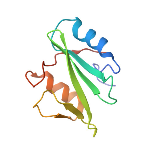
The X-ray Crystallographic Structure of Human EAT2 (SH2D1B).

Taha, M.Nezerwa, E.Nam, H.J.

(2016) Protein Pept Lett 23: 862-866

  • DOI: https://doi.org/10.2174/0929866523666160831162239
  • Primary Citation Related Structures: 
    5KAZ

  • PubMed Abstract: 

    Ewing's Sarcoma transcript-2 (EAT2) also known as SH2D1B is involved in regulation of signalling lymphocytic activation molecule (SLAM) family receptor functions. Cytoplasmic tails of SLAM family receptors contain tyrosine residues which mediate the downstream signal transduction through their phosphorylation. EAT2, composed of a single SH2 domain and a short C-terminal tail, binds to the phosphotyrosine residues and regulates SLAM family receptor signalling. We have determined the crystal structure of the human EAT2 protein in an unliganded form. Compared with the mouse EAT2-peptide complex structure, we observe conformational differences in the loops involved in ligand binding. When compared with SAP, the other single SH2 domain protein in human, EAT2 shows similar binding energies to unphosphorylated ligands. This is inconsistent to the previous data showing low affinity of EAT2 toward unphosphorylated peptides compared to SAP which shows high affinity. Additional factors other than the SH2 domains may contribute to the reported differences.


  • Organizational Affiliation
    • Department of Bioengineering, The University of Texas at Dallas, 800 W. Campbell Rd, RL10, Richardson, USA. hnam@utdallas.edu.

Macromolecule Content 

  • Total Structure Weight: 12.19 kDa 
  • Atom Count: 999 
  • Modeled Residue Count: 103 
  • Deposited Residue Count: 104 
  • Unique protein chains: 1

Macromolecules

Find similar proteins by:|  3D Structure
Entity ID: 1
MoleculeChains  Sequence LengthOrganismDetailsImage
SH2 domain-containing protein 1B104Homo sapiensMutation(s): 0 
Gene Names: SH2D1BEAT2
UniProt & NIH Common Fund Data Resources
Find proteins for O14796 (Homo sapiens)
Explore O14796 
Go to UniProtKB:  O14796
PHAROS:  O14796
GTEx:  ENSG00000198574 
Entity Groups
Sequence Clusters30% Identity50% Identity70% Identity90% Identity95% Identity100% Identity
UniProt GroupO14796
Sequence Annotations
Expand
Reference Sequence

Small Molecules

Ligands 1 Unique
IDChains Name / Formula / InChI Key2D Diagram3D Interactions
SO4

Query on SO4


Download:Ideal Coordinates CCD File
B [auth A]SULFATE ION
O4 S
QAOWNCQODCNURD-UHFFFAOYSA-L

Experimental Data & Validation

Experimental Data

  • Method: X-RAY DIFFRACTION
  • Resolution: 1.70 Å
  • R-Value Free:  0.214 (Depositor), 0.215 (DCC) 
  • R-Value Work:  0.189 (Depositor), 0.193 (DCC) 
  • R-Value Observed: 0.191 (Depositor) 
Space Group: I 4 2 2
Unit Cell:
Length ( Å )Angle ( ˚ )
a = 66.983α = 90
b = 66.983β = 90
c = 105.155γ = 90
Software Package:
Software NamePurpose
HKL-2000data reduction
HKL-2000data scaling
PHASERphasing
PHENIXrefinement

Structure Validation

View Full Validation Report


Entry History 

Deposition Data

Revision History  (Full details and data files)

  • Version 1.0: 2017-04-12
    Type: Initial release
  • Version 1.1: 2024-02-28
    Changes: Data collection, Database references