5K90 | pdb_00005k90

Crystal structure of dimeric chlorite dismutase from Cyanothece sp. PCC7425 in complex with isothiocyanate


Experimental Data Snapshot

  • Method: X-RAY DIFFRACTION
  • Resolution: 1.28 Å
  • R-Value Free: 
    0.236 (Depositor), 0.237 (DCC) 
  • R-Value Work: 
    0.210 (Depositor), 0.211 (DCC) 
  • R-Value Observed: 
    0.211 (Depositor) 

wwPDB Validation 3D Report Full Report

Validation slider image for 5K90

Ligand Structure Quality Assessment 


This is version 1.3 of the entry. See complete history

Literature

Molecular Mechanism of Enzymatic Chlorite Detoxification: Insights from Structural and Kinetic Studies.

Schaffner, I.Mlynek, G.Flego, N.Puhringer, D.Libiseller-Egger, J.Coates, L.Hofbauer, S.Bellei, M.Furtmuller, P.G.Battistuzzi, G.Smulevich, G.Djinovic-Carugo, K.Obinger, C.

(2017) ACS Catal 7: 7962-7976

  • DOI: https://doi.org/10.1021/acscatal.7b01749
  • Primary Citation Related Structures: 
    5K8Z, 5K90, 5K91, 5MAU, 5NKU, 5NKV

  • PubMed Abstract: 

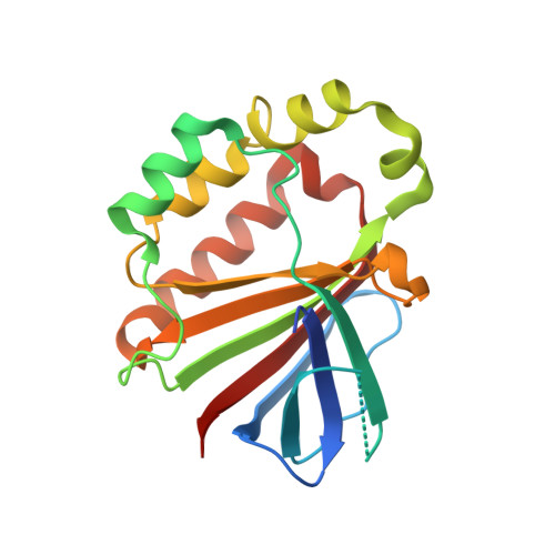
    The heme enzyme chlorite dismutase (Cld) catalyzes the degradation of chlorite to chloride and dioxygen. Although structure and steady-state kinetics of Clds have been elucidated, many questions remain (e.g., the mechanism of chlorite cleavage and the pH dependence of the reaction). Here, we present high-resolution X-ray crystal structures of a dimeric Cld at pH 6.5 and 8.5, its fluoride and isothiocyanate complexes and the neutron structure at pH 9.0 together with the pH dependence of the Fe(III)/Fe(II) couple, and the UV-vis and resonance Raman spectral features. We demonstrate that the distal Arg127 cannot act as proton acceptor and is fully ionized even at pH 9.0 ruling out its proposed role in dictating the pH dependence of chlorite degradation. Stopped-flow studies show that (i) Compound I and hypochlorite do not recombine and (ii) Compound II is the immediately formed redox intermediate that dominates during turnover. Homolytic cleavage of chlorite is proposed.


  • Organizational Affiliation
    • Department of Chemistry, Division of Biochemistry, BOKU-University of Natural Resources and Life Sciences, Muthgasse 18, A-1190 Vienna, Austria.

Macromolecule Content 

  • Total Structure Weight: 91.07 kDa 
  • Atom Count: 6,866 
  • Modeled Residue Count: 698 
  • Deposited Residue Count: 752 
  • Unique protein chains: 1

Macromolecules

Find similar proteins by:|  3D Structure
Entity ID: 1
MoleculeChains  Sequence LengthOrganismDetailsImage
Chlorite dismutase
A, B, C, D
188Cyanothece sp. PCC 7425Mutation(s): 0 
Gene Names: Cyan7425_1434
EC: 1.3.98.5
UniProt
Find proteins for B8HNS6 (Cyanothece sp. (strain PCC 7425 / ATCC 29141))
Explore B8HNS6 
Go to UniProtKB:  B8HNS6
Entity Groups
Sequence Clusters30% Identity50% Identity70% Identity90% Identity95% Identity100% Identity
UniProt GroupB8HNS6
Sequence Annotations
Expand
Reference Sequence

Small Molecules

Ligands 5 Unique
IDChains Name / Formula / InChI Key2D Diagram3D Interactions
HEM

Query on HEM


Download:Ideal Coordinates CCD File
F [auth A],
I [auth B],
O [auth C],
V [auth D]
PROTOPORPHYRIN IX CONTAINING FE
C34 H32 Fe N4 O4
KABFMIBPWCXCRK-RGGAHWMASA-L
SO4

Query on SO4


Download:Ideal Coordinates CCD File
H [auth A],
T [auth C]
SULFATE ION
O4 S
QAOWNCQODCNURD-UHFFFAOYSA-L
GOL

Query on GOL


Download:Ideal Coordinates CCD File
G [auth A]
K [auth B]
L [auth B]
M [auth B]
P [auth C]
G [auth A],
K [auth B],
L [auth B],
M [auth B],
P [auth C],
Q [auth C],
R [auth C],
S [auth C],
X [auth D]
GLYCEROL
C3 H8 O3
PEDCQBHIVMGVHV-UHFFFAOYSA-N
IS8

Query on IS8


Download:Ideal Coordinates CCD File
E [auth A],
J [auth B],
N [auth C],
W [auth D]
isothiocyanate
C H N S
GRHBQAYDJPGGLF-UHFFFAOYSA-N
MG

Query on MG


Download:Ideal Coordinates CCD File
U [auth C]MAGNESIUM ION
Mg
JLVVSXFLKOJNIY-UHFFFAOYSA-N

Experimental Data & Validation

Experimental Data

  • Method: X-RAY DIFFRACTION
  • Resolution: 1.28 Å
  • R-Value Free:  0.236 (Depositor), 0.237 (DCC) 
  • R-Value Work:  0.210 (Depositor), 0.211 (DCC) 
  • R-Value Observed: 0.211 (Depositor) 
Space Group: P 1
Unit Cell:
Length ( Å )Angle ( ˚ )
a = 51.759α = 99.8
b = 54.631β = 94.82
c = 94.096γ = 99.17
Software Package:
Software NamePurpose
PHENIXrefinement
XDSdata reduction
XDSdata scaling
PHENIXphasing

Structure Validation

View Full Validation Report


Ligand Structure Quality Assessment 


Entry History 

Deposition Data

Revision History  (Full details and data files)

  • Version 1.0: 2017-06-21
    Type: Initial release
  • Version 1.1: 2018-01-31
    Changes: Database references
  • Version 1.2: 2019-10-02
    Changes: Data collection, Derived calculations
  • Version 1.3: 2024-05-08
    Changes: Data collection, Database references, Derived calculations