5K7H | pdb_00005k7h

Crystal structure of AibR in complex with the effector molecule isovaleryl coenzyme A


Experimental Data Snapshot

  • Method: X-RAY DIFFRACTION
  • Resolution: 2.35 Å
  • R-Value Free: 
    0.235 (Depositor), 0.238 (DCC) 
  • R-Value Work: 
    0.192 (Depositor), 0.197 (DCC) 
  • R-Value Observed: 
    0.194 (Depositor) 

Starting Model: experimental
View more details

wwPDB Validation 3D Report Full Report

Validation slider image for 5K7H

Ligand Structure Quality Assessment 


This is version 1.2 of the entry. See complete history

Literature

The AibR-isovaleryl coenzyme A regulator and its DNA binding site - a model for the regulation of alternative de novo isovaleryl coenzyme A biosynthesis in Myxococcus xanthus.

Bock, T.Volz, C.Hering, V.Scrima, A.Muller, R.Blankenfeldt, W.

(2017) Nucleic Acids Res 45: 2166-2178

  • DOI: https://doi.org/10.1093/nar/gkw1238
  • Primary Citation Related Structures: 
    5K7F, 5K7H

  • PubMed Abstract: 

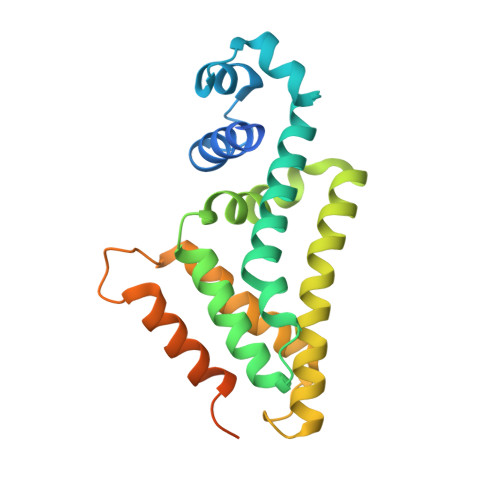
    Isovaleryl coenzyme A (IV-CoA) is an important building block of iso-fatty acids. In myxobacteria, IV-CoA is essential for the formation of signaling molecules involved in fruiting body formation. Leucine degradation is the common source of IV-CoA, but a second, de novo biosynthetic route to IV-CoA termed AIB (alternative IV-CoA biosynthesis) was recently discovered in M. xanthus. The AIB-operon contains the TetR-like transcriptional regulator AibR, which we characterize in this study. We demonstrate that IV-CoA binds AibR with micromolar affinity and show by gelshift experiments that AibR interacts with the promoter region of the AIB-operon once IV-CoA is present. We identify an 18-bp near-perfect palindromic repeat as containing the AibR operator and provide evidence that AibR also controls an additional genomic locus coding for a putative acetyl-CoA acetyltransferase. To elucidate atomic details, we determined crystal structures of AibR in the apo, the IV-CoA- and the IV-CoA-DNA-bound state to 1.7 Å, 2.35 Å and 2.92 Å, respectively. IV-CoA induces partial unfolding of an α-helix, which allows sequence-specific interactions between AibR and its operator. This study provides insights into AibR-mediated regulation and shows that AibR functions in an unusual TetR-like manner by blocking transcription not in the ligand-free but in the effector-bound state.


  • Organizational Affiliation
    • Structure and Function of Proteins, Helmholtz Centre for Infection Research, Inhoffenstr. 7, 38124 Braunschweig, Germany.

Macromolecule Content 

  • Total Structure Weight: 53.27 kDa 
  • Atom Count: 3,022 
  • Modeled Residue Count: 368 
  • Deposited Residue Count: 462 
  • Unique protein chains: 1

Macromolecules

Find similar proteins by:|  3D Structure
Entity ID: 1
MoleculeChains  Sequence LengthOrganismDetailsImage
Transcriptional regulator, TetR family
A, B
231Myxococcus xanthus DK 1622Mutation(s): 0 
Gene Names: MXAN_4263
UniProt
Find proteins for Q1D4I5 (Myxococcus xanthus (strain DK1622))
Explore Q1D4I5 
Go to UniProtKB:  Q1D4I5
Entity Groups
Sequence Clusters30% Identity50% Identity70% Identity90% Identity95% Identity100% Identity
UniProt GroupQ1D4I5
Sequence Annotations
Expand
Reference Sequence

Small Molecules

Ligands 3 Unique
IDChains Name / Formula / InChI Key2D Diagram3D Interactions
IVC

Query on IVC


Download:Ideal Coordinates CCD File
G [auth A],
I [auth B]
Isovaleryl-coenzyme A
C26 H44 N7 O17 P3 S
UYVZIWWBJMYRCD-ZMHDXICWSA-N
NI

Query on NI


Download:Ideal Coordinates CCD File
C [auth A]NICKEL (II) ION
Ni
VEQPNABPJHWNSG-UHFFFAOYSA-N
CL

Query on CL


Download:Ideal Coordinates CCD File
D [auth A],
E [auth A],
F [auth A],
H [auth B]
CHLORIDE ION
Cl
VEXZGXHMUGYJMC-UHFFFAOYSA-M

Experimental Data & Validation

Experimental Data

  • Method: X-RAY DIFFRACTION
  • Resolution: 2.35 Å
  • R-Value Free:  0.235 (Depositor), 0.238 (DCC) 
  • R-Value Work:  0.192 (Depositor), 0.197 (DCC) 
  • R-Value Observed: 0.194 (Depositor) 
Space Group: P 61
Unit Cell:
Length ( Å )Angle ( ˚ )
a = 89.78α = 90
b = 89.78β = 90
c = 132.118γ = 120
Software Package:
Software NamePurpose
PHENIXrefinement
Aimlessdata scaling
PDB_EXTRACTdata extraction
XDSdata reduction
PHENIXphasing

Structure Validation

View Full Validation Report


Ligand Structure Quality Assessment 


Entry History 

Deposition Data

Revision History  (Full details and data files)

  • Version 1.0: 2016-12-21
    Type: Initial release
  • Version 1.1: 2017-05-03
    Changes: Database references
  • Version 1.2: 2024-01-10
    Changes: Data collection, Database references, Derived calculations, Refinement description