Structural basis for dual specificity of yeast N-terminal amidase in the N-end rule pathway.
Kim, M.K., Oh, S.J., Lee, B.G., Song, H.K.(2016) Proc Natl Acad Sci U S A 113: 12438-12443
- PubMed: 27791147 Search on PubMedSearch on PubMed Central
- DOI: https://doi.org/10.1073/pnas.1612620113
- Primary Citation Related Structures:
5B62, 5HYY, 5K5U, 5K5V, 5K60, 5K61, 5K62, 5K63, 5K66 - PubMed Abstract:
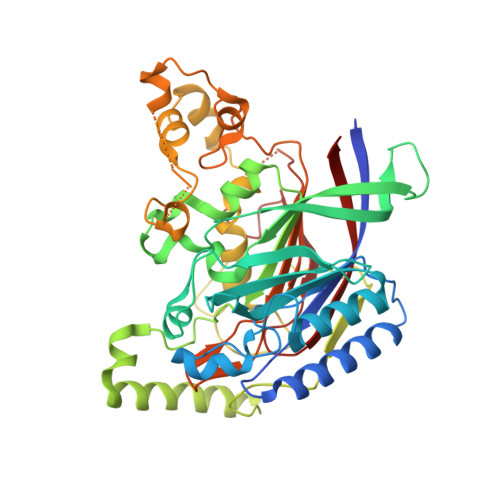
The first step of the hierarchically organized Arg/N-end rule pathway of protein degradation is deamidation of the N-terminal glutamine and asparagine residues of substrate proteins to glutamate and aspartate, respectively. These reactions are catalyzed by the N-terminal amidase (Nt-amidase) Nta1 in fungi such as Saccharomyces cerevisiae, and by the glutamine-specific Ntaq1 and asparagine-specific Ntan1 Nt-amidases in mammals. To investigate the dual specificity of yeast Nta1 (yNta1) and the importance of second-position residues in Asn/Gln-bearing N-terminal degradation signals (N-degrons), we determined crystal structures of yNta1 in the apo state and in complex with various N-degron peptides. Both an Asn-peptide and a Gln-peptide fit well into the hollow active site pocket of yNta1, with the catalytic triad located deeper inside the active site. Specific hydrogen bonds stabilize interactions between N-degron peptides and hydrophobic peripheral regions of the active site pocket. Key determinants for substrate recognition were identified and thereafter confirmed by using structure-based mutagenesis. We also measured affinities between yNta1 (wild-type and its mutants) and specific peptides, and determined K M and k cat for peptides of each type. Together, these results elucidate, in structural and mechanistic detail, specific deamidation mechanisms in the first step of the N-end rule pathway.
- Department of Life Sciences, Korea University, Seoul 02841, Korea.
Organizational Affiliation: