5K5N | pdb_00005k5n

Crystal structure of GSK-3beta complexed with PF-04802367, a highly selective brain-penetrant kinase inhibitor


Experimental Data Snapshot

  • Method: X-RAY DIFFRACTION
  • Resolution: 2.20 Å
  • R-Value Free: 
    0.229 (Depositor), 0.230 (DCC) 
  • R-Value Work: 
    0.204 (Depositor), 0.203 (DCC) 
  • R-Value Observed: 
    0.205 (Depositor) 

wwPDB Validation 3D Report Full Report

Validation slider image for 5K5N

Ligand Structure Quality Assessment 


This is version 1.1 of the entry. See complete history

Literature

Discovery of a Highly Selective Glycogen Synthase Kinase-3 Inhibitor (PF-04802367) That Modulates Tau Phosphorylation in the Brain: Translation for PET Neuroimaging.

Liang, S.H.Chen, J.M.Normandin, M.D.Chang, J.S.Chang, G.C.Taylor, C.K.Trapa, P.Plummer, M.S.Para, K.S.Conn, E.L.Lopresti-Morrow, L.Lanyon, L.F.Cook, J.M.Richter, K.E.Nolan, C.E.Schachter, J.B.Janat, F.Che, Y.Shanmugasundaram, V.Lefker, B.A.Enerson, B.E.Livni, E.Wang, L.Guehl, N.J.Patnaik, D.Wagner, F.F.Perlis, R.Holson, E.B.Haggarty, S.J.El Fakhri, G.Kurumbail, R.G.Vasdev, N.

(2016) Angew Chem Int Ed Engl 55: 9601-9605

  • DOI: https://doi.org/10.1002/anie.201603797
  • Primary Citation Related Structures: 
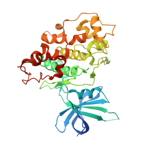
    5K5N

  • PubMed Abstract: 

    Glycogen synthase kinase-3 (GSK-3) regulates multiple cellular processes in diabetes, oncology, and neurology. N-(3-(1H-1,2,4-triazol-1-yl)propyl)-5-(3-chloro-4-methoxyphenyl)oxazole-4-carboxamide (PF-04802367 or PF-367) has been identified as a highly potent inhibitor, which is among the most selective antagonists of GSK-3 to date. Its efficacy was demonstrated in modulation of tau phosphorylation in vitro and in vivo. Whereas the kinetics of PF-367 binding in brain tissues are too fast for an effective therapeutic agent, the pharmacokinetic profile of PF-367 is ideal for discovery of radiopharmaceuticals for GSK-3 in the central nervous system. A (11) C-isotopologue of PF-367 was synthesized and preliminary PET imaging studies in non-human primates confirmed that we have overcome the two major obstacles for imaging GSK-3, namely, reasonable brain permeability and displaceable binding.


  • Organizational Affiliation
    • Gordon Center for Medical Imaging & Nuclear Medicine and Molecular Imaging, Massachusetts General Hospital & Department of Radiology, Harvard Medical School, Boston, MA, 02114, USA.

Macromolecule Content 

  • Total Structure Weight: 85.17 kDa 
  • Atom Count: 5,484 
  • Modeled Residue Count: 667 
  • Deposited Residue Count: 740 
  • Unique protein chains: 1

Macromolecules

Find similar proteins by:|  3D Structure
Entity ID: 1
MoleculeChains  Sequence LengthOrganismDetailsImage
Glycogen synthase kinase-3 beta
A, B
370Homo sapiensMutation(s): 1 
Gene Names: GSK3B
EC: 2.7.11.26 (PDB Primary Data), 2.7.11.1 (PDB Primary Data)
UniProt & NIH Common Fund Data Resources
Find proteins for P49841 (Homo sapiens)
Explore P49841 
Go to UniProtKB:  P49841
PHAROS:  P49841
GTEx:  ENSG00000082701 
Entity Groups
Sequence Clusters30% Identity50% Identity70% Identity90% Identity95% Identity100% Identity
UniProt GroupP49841
Sequence Annotations
Expand
Reference Sequence

Small Molecules

Experimental Data & Validation

Experimental Data

  • Method: X-RAY DIFFRACTION
  • Resolution: 2.20 Å
  • R-Value Free:  0.229 (Depositor), 0.230 (DCC) 
  • R-Value Work:  0.204 (Depositor), 0.203 (DCC) 
  • R-Value Observed: 0.205 (Depositor) 
Space Group: P 1 21 1
Unit Cell:
Length ( Å )Angle ( ˚ )
a = 66.293α = 90
b = 99.907β = 102.9
c = 66.91γ = 90
Software Package:
Software NamePurpose
BUSTERrefinement
DENZOdata reduction
SCALEPACKdata scaling
PDB_EXTRACTdata extraction
REFMACphasing

Structure Validation

View Full Validation Report


Ligand Structure Quality Assessment 


Entry History 

Deposition Data

Revision History  (Full details and data files)

  • Version 1.0: 2016-09-21
    Type: Initial release
  • Version 1.1: 2024-11-20
    Changes: Data collection, Database references, Derived calculations, Structure summary