5K1H

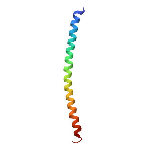
eIF3b relocated to the intersubunit face to interact with eIF1 and below the eIF2 ternary-complex. from the structure of a partial yeast 48S preinitiation complex in closed conformation.


Experimental Data Snapshot

  • Method: ELECTRON MICROSCOPY
  • Resolution: 4.90 Å
  • Aggregation State: PARTICLE 
  • Reconstruction Method: SINGLE PARTICLE 

Starting Model: experimental
View more details

wwPDB Validation   3D Report Full Report


This is version 2.2 of the entry. See complete history


Literature

eIF3 Peripheral Subunits Rearrangement after mRNA Binding and Start-Codon Recognition.

Simonetti, A.Brito Querido, J.Myasnikov, A.G.Mancera-Martinez, E.Renaud, A.Kuhn, L.Hashem, Y.

(2016) Mol Cell 63: 206-217

  • DOI: https://doi.org/10.1016/j.molcel.2016.05.033
  • Primary Citation of Related Structures:  
    5K0Y, 5K1H

  • PubMed Abstract: 

    mRNA translation initiation in eukaryotes requires the cooperation of a dozen eukaryotic initiation factors (eIFs) forming several complexes, which leads to mRNA attachment to the small ribosomal 40S subunit, mRNA scanning for start codon, and accommodation of initiator tRNA at the 40S P site. eIF3, composed of 13 subunits, 8 core (a, c, e, f, h, l, k, and m) and 5 peripheral (b, d, g, i, and j), plays a central role during this process. Here we report a cryo-electron microscopy structure of a mammalian 48S initiation complex at 5.8 Å resolution. It shows the relocation of subunits eIF3i and eIF3g to the 40S intersubunit face on the GTPase binding site, at a late stage in initiation. On the basis of a previous study, we demonstrate the relocation of eIF3b to the 40S intersubunit face, binding below the eIF2-Met-tRNAi(Met) ternary complex upon mRNA attachment. Our analysis reveals the deep rearrangement of eIF3 and unravels the molecular mechanism underlying eIF3 function in mRNA scanning and timing of ribosomal subunit joining.


  • Organizational Affiliation

    CNRS, Architecture et Réactivité de l'ARN UPR9002, Université de Strasbourg, 67084 Strasbourg, France. Electronic address: a.simonetti@ibmc-cnrs.unistra.fr.


Macromolecules
Find similar proteins by:  (by identity cutoff)  |  3D Structure
Entity ID: 1
MoleculeChains Sequence LengthOrganismDetailsImage
Eukaryotic translation initiation factor 3 subunit BA [auth B]576Homo sapiensMutation(s): 0 
Gene Names: EIF3BEIF3S9
UniProt & NIH Common Fund Data Resources
Find proteins for P55884 (Homo sapiens)
Explore P55884 
Go to UniProtKB:  P55884
PHAROS:  P55884
GTEx:  ENSG00000106263 
Entity Groups  
Sequence Clusters30% Identity50% Identity70% Identity90% Identity95% Identity100% Identity
UniProt GroupP55884
Sequence Annotations
Expand
  • Reference Sequence
Find similar proteins by:  (by identity cutoff)  |  3D Structure
Entity ID: 2
MoleculeChains Sequence LengthOrganismDetailsImage
eIF3a C-terminal tailB [auth A]54Oryctolagus cuniculusMutation(s): 0 
Sequence Annotations
Expand
  • Reference Sequence
Experimental Data & Validation

Experimental Data

  • Method: ELECTRON MICROSCOPY
  • Resolution: 4.90 Å
  • Aggregation State: PARTICLE 
  • Reconstruction Method: SINGLE PARTICLE 
EM Software:
TaskSoftware PackageVersion
MODEL REFINEMENTVMD

Structure Validation

View Full Validation Report


Entry History 

Deposition Data

Revision History  (Full details and data files)

  • Version 1.0: 2016-07-13
    Type: Initial release
  • Version 1.1: 2016-08-03
    Changes: Database references
  • Version 1.2: 2017-08-02
    Changes: Data collection
  • Version 2.0: 2017-08-09
    Changes: Advisory, Atomic model, Database references, Derived calculations, Polymer sequence, Source and taxonomy, Structure summary
  • Version 2.1: 2018-01-31
    Changes: Database references
  • Version 2.2: 2024-05-08
    Changes: Data collection, Database references, Refinement description