5K01 | pdb_00005k01

Crystal Structure of COMT in complex with 2,7-dimethyl-3-(1H-pyrazol-3-yl)imidazo[1,2-a]pyridine


Experimental Data Snapshot

  • Method: X-RAY DIFFRACTION
  • Resolution: 1.38 Å
  • R-Value Free: 
    0.176 (Depositor), 0.179 (DCC) 
  • R-Value Work: 
    0.134 (Depositor), 0.137 (DCC) 
  • R-Value Observed: 
    0.137 (Depositor) 

wwPDB Validation 3D Report Full Report

Validation slider image for 5K01

Ligand Structure Quality Assessment 


This is version 1.2 of the entry. See complete history

Literature

Crystal Structure of COMT

Ehler, A.Rodriguez-Sarmiento, R.M.Rudolph, M.G.

To be published.

Macromolecule Content 

  • Total Structure Weight: 24.24 kDa 
  • Atom Count: 2,057 
  • Modeled Residue Count: 214 
  • Deposited Residue Count: 214 
  • Unique protein chains: 1

Macromolecules

Find similar proteins by:|  3D Structure
Entity ID: 1
MoleculeChains  Sequence LengthOrganismDetailsImage
Catechol O-methyltransferase214Rattus norvegicusMutation(s): 0 
Gene Names: Comt
EC: 2.1.1.6
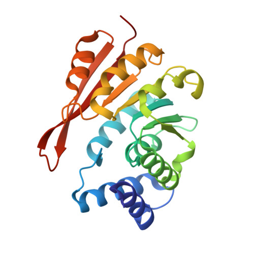
UniProt
Find proteins for P22734 (Rattus norvegicus)
Explore P22734 
Go to UniProtKB:  P22734
Entity Groups
Sequence Clusters30% Identity50% Identity70% Identity90% Identity95% Identity100% Identity
UniProt GroupP22734
Sequence Annotations
Expand
Reference Sequence

Small Molecules

Ligands 2 Unique
IDChains Name / Formula / InChI Key2D Diagram3D Interactions
6OW

Query on 6OW


Download:Ideal Coordinates CCD File
B [auth A]2,7-dimethyl-3-(1H-pyrazol-5-yl)imidazo[1,2-a]pyridine
C12 H12 N4
ZNRUSTFXUSPVPD-UHFFFAOYSA-N
K

Query on K


Download:Ideal Coordinates CCD File
C [auth A]POTASSIUM ION
K
NPYPAHLBTDXSSS-UHFFFAOYSA-N

Experimental Data & Validation

Experimental Data

  • Method: X-RAY DIFFRACTION
  • Resolution: 1.38 Å
  • R-Value Free:  0.176 (Depositor), 0.179 (DCC) 
  • R-Value Work:  0.134 (Depositor), 0.137 (DCC) 
  • R-Value Observed: 0.137 (Depositor) 
Space Group: P 21 21 21
Unit Cell:
Length ( Å )Angle ( ˚ )
a = 33.789α = 90
b = 59.788β = 90
c = 108.093γ = 90
Software Package:
Software NamePurpose
XSCALEdata scaling
PDB_EXTRACTdata extraction
PHENIXrefinement
XDSdata reduction
PHENIXphasing

Structure Validation

View Full Validation Report


Ligand Structure Quality Assessment 


Entry History 

Deposition Data

Revision History  (Full details and data files)

  • Version 1.0: 2016-09-07
    Type: Initial release
  • Version 1.1: 2019-06-12
    Changes: Data collection, Database references, Structure summary
  • Version 1.2: 2024-05-08
    Changes: Data collection, Database references