5JX2 | pdb_00005jx2

Crystal structure of MglB-2 (Tp0684) from Treponema pallidum


Experimental Data Snapshot

  • Method: X-RAY DIFFRACTION
  • Resolution: 2.05 Å
  • R-Value Free: 
    0.226 (Depositor), 0.233 (DCC) 
  • R-Value Work: 
    0.186 (Depositor), 0.195 (DCC) 
  • R-Value Observed: 
    0.188 (Depositor) 

wwPDB Validation 3D Report Full Report

Validation slider image for 5JX2

This is version 2.1 of the entry. See complete history

Literature

The Tp0684 (MglB-2) Lipoprotein of Treponema pallidum: A Glucose-Binding Protein with Divergent Topology.

Brautigam, C.A.Deka, R.K.Liu, W.Z.Norgard, M.V.

(2016) PLoS One 11: e0161022-e0161022

  • DOI: https://doi.org/10.1371/journal.pone.0161022
  • Primary Citation Related Structures: 
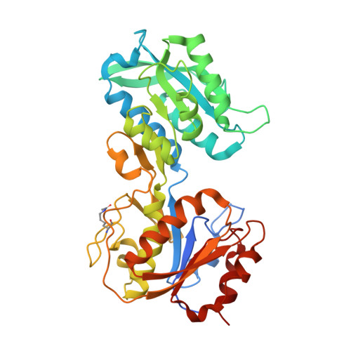
    5JX2

  • PubMed Abstract: 

    Treponema pallidum, the bacterium that causes syphilis, is an obligate human parasite. As such, it must acquire energy, in the form of carbon sources, from the host. There is ample evidence that the principal source of energy for this spirochete is D-glucose acquired from its environment, likely via an ABC transporter. Further, there is genetic evidence of a D-glucose chemotaxis system in T. pallidum. Both of these processes may be dependent on a single lipidated chemoreceptor: Tp0684, also called TpMglB-2 for its sequence homology to MglB of Escherichia coli. To broaden our understanding of this potentially vital protein, we determined a 2.05-Å X-ray crystal structure of a soluble form of the recombinant protein. Like its namesake, TpMglB-2 adopts a bilobed fold that is similar to that of the ligand-binding proteins (LBPs) of other ABC transporters. However, the protein has an unusual, circularly permuted topology. This feature prompted a series of biophysical studies that examined whether the protein's topological distinctiveness affected its putative chemoreceptor functions. Differential scanning fluorimetry and isothermal titration calorimetry were used to confirm that the protein bound D-glucose in a cleft between its two lobes. Additionally, analytical ultracentrifugation was employed to reveal that D-glucose binding is accompanied by a significant conformational change. TpMglB-2 thus appears to be fully functional in vitro, and given the probable central importance of the protein to T. pallidum's physiology, our results have implications for the viability and pathogenicity of this obligate human pathogen.


  • Organizational Affiliation
    • Department of Biophysics, The University of Texas Southwestern Medical Center, Dallas, TX, 75390, United States of America.

Macromolecule Content 

  • Total Structure Weight: 42.19 kDa 
  • Atom Count: 2,992 
  • Modeled Residue Count: 366 
  • Deposited Residue Count: 387 
  • Unique protein chains: 1

Macromolecules

Find similar proteins by:|  3D Structure
Entity ID: 1
MoleculeChains  Sequence LengthOrganismDetailsImage
Glucose/galactose-binding lipoprotein387Treponema pallidum subsp. pallidum str. NicholsMutation(s): 0 
Gene Names: mglBtpp38TP_0684
UniProt
Find proteins for Q08255 (Treponema pallidum (strain Nichols))
Explore Q08255 
Go to UniProtKB:  Q08255
Entity Groups
Sequence Clusters30% Identity50% Identity70% Identity90% Identity95% Identity100% Identity
UniProt GroupQ08255
Sequence Annotations
Expand
Reference Sequence

Experimental Data & Validation

Experimental Data

  • Method: X-RAY DIFFRACTION
  • Resolution: 2.05 Å
  • R-Value Free:  0.226 (Depositor), 0.233 (DCC) 
  • R-Value Work:  0.186 (Depositor), 0.195 (DCC) 
  • R-Value Observed: 0.188 (Depositor) 
Space Group: C 2 2 21
Unit Cell:
Length ( Å )Angle ( ˚ )
a = 77.556α = 90
b = 130.025β = 90
c = 88.25γ = 90
Software Package:
Software NamePurpose
PHENIXrefinement
HKL-3000data reduction
HKL-3000data scaling
HKL-3000phasing

Structure Validation

View Full Validation Report


Entry History 

& Funding Information

Deposition Data


Funding OrganizationLocationGrant Number
National Institutes of Health/National Institute Of Allergy and Infectious Diseases (NIH/NIAID)United StatesAI056305

Revision History  (Full details and data files)

  • Version 1.0: 2016-08-31
    Type: Initial release
  • Version 1.1: 2017-09-06
    Changes: Author supporting evidence, Derived calculations
  • Version 1.2: 2019-12-11
    Changes: Author supporting evidence
  • Version 2.0: 2023-11-15
    Changes: Atomic model, Data collection, Database references, Derived calculations
  • Version 2.1: 2024-11-06
    Changes: Structure summary