Bacterial protease uses distinct thermodynamic signatures for substrate recognition.
Bezerra, G.A., Ohara-Nemoto, Y., Cornaciu, I., Fedosyuk, S., Hoffmann, G., Round, A., Marquez, J.A., Nemoto, T.K., Djinovic-Carugo, K.(2017) Sci Rep 7: 2848-2848
- PubMed: 28588213
- DOI: https://doi.org/10.1038/s41598-017-03220-y
- Primary Citation of Related Structures:
5JWF, 5JWG, 5JWI, 5JXF, 5JXK, 5JXP, 5JY0 - PubMed Abstract:
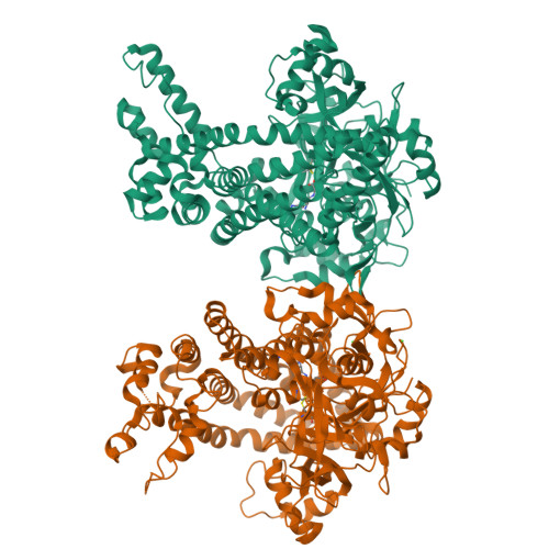
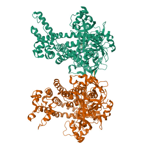
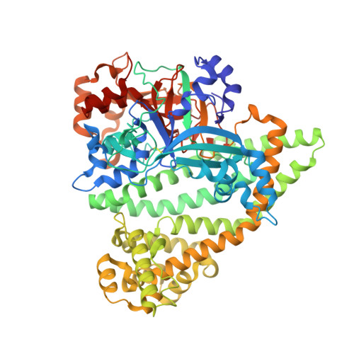
Porphyromonas gingivalis and Porphyromonas endodontalis are important bacteria related to periodontitis, the most common chronic inflammatory disease in humans worldwide. Its comorbidity with systemic diseases, such as type 2 diabetes, oral cancers and cardiovascular diseases, continues to generate considerable interest. Surprisingly, these two microorganisms do not ferment carbohydrates; rather they use proteinaceous substrates as carbon and energy sources. However, the underlying biochemical mechanisms of their energy metabolism remain unknown. Here, we show that dipeptidyl peptidase 11 (DPP11), a central metabolic enzyme in these bacteria, undergoes a conformational change upon peptide binding to distinguish substrates from end products. It binds substrates through an entropy-driven process and end products in an enthalpy-driven fashion. We show that increase in protein conformational entropy is the main-driving force for substrate binding via the unfolding of specific regions of the enzyme ("entropy reservoirs"). The relationship between our structural and thermodynamics data yields a distinct model for protein-protein interactions where protein conformational entropy modulates the binding free-energy. Further, our findings provide a framework for the structure-based design of specific DPP11 inhibitors.
Organizational Affiliation:
Department of Structural & Computational Biology, Max F. Perutz Laboratories, University of Vienna, Vienna Biocenter, Vienna Biocenter Campus 5, A-1030, Vienna, Austria. gustavo.bezerra@univie.ac.at.


