5JUG | pdb_00005jug

Structure of an inactive (E45Q) variant of a beta-1,4-mannanase, SsGH134, in complex with Man5


Experimental Data Snapshot

  • Method: X-RAY DIFFRACTION
  • Resolution: 0.96 Å
  • R-Value Free: 
    0.141 (Depositor), 0.122 (DCC) 
  • R-Value Work: 
    0.122 (Depositor) 
  • R-Value Observed: 
    0.123 (Depositor) 

Starting Model: other
View more details

wwPDB Validation 3D Report Full Report

Validation slider image for 5JUG

Ligand Structure Quality Assessment 


This is version 2.2 of the entry. See complete history

Literature

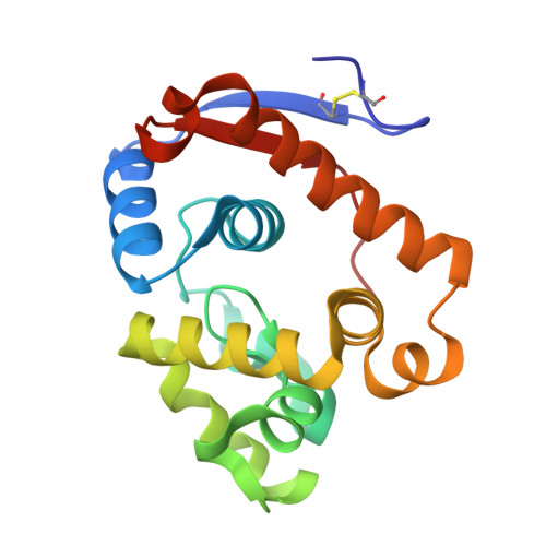
A beta-Mannanase with a Lysozyme-like Fold and a Novel Molecular Catalytic Mechanism.

Jin, Y.Petricevic, M.John, A.Raich, L.Jenkins, H.Portela De Souza, L.Cuskin, F.Gilbert, H.J.Rovira, C.Goddard-Borger, E.D.Williams, S.J.Davies, G.J.

(2016) ACS Cent Sci 2: 896-903

  • DOI: https://doi.org/10.1021/acscentsci.6b00232
  • Primary Citation Related Structures: 
    5JTS, 5JU9, 5JUG

  • PubMed Abstract: 

    The enzymatic cleavage of β-1,4-mannans is achieved by endo -β-1,4-mannanases, enzymes involved in germination of seeds and microbial hemicellulose degradation, and which have increasing industrial and consumer product applications. β-Mannanases occur in a range of families of the CAZy sequence-based glycoside hydrolase (GH) classification scheme including families 5, 26, and 113. In this work we reveal that β-mannanases of the newly described GH family 134 differ from other mannanase families in both their mechanism and tertiary structure. A representative GH family 134 endo -β-1,4-mannanase from a Streptomyces sp. displays a fold closely related to that of hen egg white lysozyme but acts with inversion of stereochemistry. A Michaelis complex with mannopentaose, and a product complex with mannotriose, reveal ligands with pyranose rings distorted in an unusual inverted chair conformation. Ab initio quantum mechanics/molecular mechanics metadynamics quantified the energetically accessible ring conformations and provided evidence in support of a 1 C 4 3 H 4 3 S 1 conformational itinerary along the reaction coordinate. This work, in concert with that on GH family 124 cellulases, reveals how the lysozyme fold can be co-opted to catalyze the hydrolysis of different polysaccharides in a mechanistically distinct manner.


  • Organizational Affiliation
    • York Structural Biology Laboratory, Department of Chemistry, University of York , Heslington, YO10 5DD, U.K.

Macromolecule Content 

  • Total Structure Weight: 18.73 kDa 
  • Atom Count: 1,581 
  • Modeled Residue Count: 168 
  • Deposited Residue Count: 168 
  • Unique protein chains: 1

Macromolecules

Find similar proteins by:|  3D Structure
Entity ID: 1
MoleculeChains  Sequence LengthOrganismDetailsImage
beta-1,4-mannanase168Streptomyces sp. NRRL B-16215Mutation(s): 0 
EC: 3.2.1.78
UniProt
Find proteins for A0ACD6BAU0 (Streptomyces sp. NRRL B-16215)
Explore A0ACD6BAU0 
Go to UniProtKB:  A0ACD6BAU0
Entity Groups
Sequence Clusters30% Identity50% Identity70% Identity90% Identity95% Identity100% Identity
UniProt GroupA0ACD6BAU0
Sequence Annotations
Expand
Reference Sequence

Oligosaccharides

Help  
Entity ID: 2
MoleculeChains Length2D Diagram GlycosylationD Interactions
beta-D-mannopyranose-(1-4)-beta-D-mannopyranose-(1-4)-beta-D-mannopyranose-(1-4)-beta-D-mannopyranose-(1-4)-beta-D-mannopyranose
B
5N/A
Glycosylation Resources
GlyTouCan: G91924WY
GlyCosmos: G91924WY
GlyGen: G91924WY

Experimental Data & Validation

Experimental Data

  • Method: X-RAY DIFFRACTION
  • Resolution: 0.96 Å
  • R-Value Free:  0.141 (Depositor), 0.122 (DCC) 
  • R-Value Work:  0.122 (Depositor) 
  • R-Value Observed: 0.123 (Depositor) 
Space Group: P 21 21 21
Unit Cell:
Length ( Å )Angle ( ˚ )
a = 36.707α = 90
b = 53.841β = 90
c = 82.636γ = 90
Software Package:
Software NamePurpose
REFMACrefinement
xia2data reduction
xia2data scaling
ACORNphasing

Structure Validation

View Full Validation Report


Ligand Structure Quality Assessment 


Entry History 

& Funding Information

Deposition Data


Funding OrganizationLocationGrant Number
European Research CouncilUnited KingdomAdG-322942
Australian Research CouncilAustralia--

Revision History  (Full details and data files)

  • Version 1.0: 2016-11-16
    Type: Initial release
  • Version 1.1: 2017-01-18
    Changes: Database references
  • Version 2.0: 2020-07-29
    Type: Remediation
    Reason: Carbohydrate remediation
    Changes: Atomic model, Data collection, Derived calculations, Structure summary
  • Version 2.1: 2024-05-01
    Changes: Data collection, Database references, Derived calculations, Refinement description, Structure summary
  • Version 2.2: 2024-10-16
    Changes: Structure summary