5JUE | pdb_00005jue

Crystal Structure of UIC2 Fab


Experimental Data Snapshot

  • Method: X-RAY DIFFRACTION
  • Resolution: 1.65 Å
  • R-Value Free: 
    0.203 (Depositor), 0.206 (DCC) 
  • R-Value Work: 
    0.165 (Depositor), 0.168 (DCC) 
  • R-Value Observed: 
    0.166 (Depositor) 

Starting Model: experimental
View more details

wwPDB Validation 3D Report Full Report

Validation slider image for 5JUE

This is version 1.2 of the entry. See complete history

Literature

Crystal structure of the antigen-binding fragment of a monoclonal antibody specific for the multidrug-resistance-linked ABC transporter human P-glycoprotein.

Esser, L.Shukla, S.Zhou, F.Ambudkar, S.V.Xia, D.

(2016) Acta Crystallogr F Struct Biol Commun 72: 636-641

  • DOI: https://doi.org/10.1107/S2053230X16009778
  • Primary Citation Related Structures: 
    5JUE

  • PubMed Abstract: 

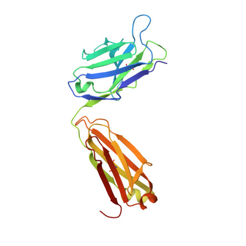
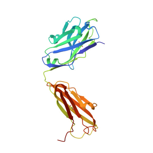
    P-glycoprotein (P-gp) is a polyspecific ATP-dependent transporter linked to multidrug resistance in cancers that plays important roles in the pharmacokinetics of a large number of drugs. The drug-resistance phenotype of P-gp can be modulated by the monoclonal antibody UIC2, which specifically recognizes human P-gp in a conformation-dependent manner. Here, the purification, sequence determination and high-resolution structure of the Fab fragment of UIC2 (UIC2/Fab) are reported. Purified UIC2/Fab binds human P-gp with a 1:1 stoichiometry. Crystals of UIC2/Fab are triclinic (space group P1), with unit-cell parameters a = 40.67, b = 44.91, c = 58.09 Å, α = 97.62, β = 99.10, γ = 94.09°, and diffracted X-rays to 1.6 Å resolution. The structure was determined by molecular replacement and refined to 1.65 Å resolution. The asymmetric unit contains one molecule of UIC2/Fab, which exhibits a positively charged antigen-binding surface, suggesting that it might recognize an oppositely charged extracellular epitope of P-gp.


  • Organizational Affiliation
    • National Cancer Institute, National Institutes of Health, 37 Convent Drive, Building 37, Bethesda, MD 20892, USA.

Macromolecule Content 

  • Total Structure Weight: 49.03 kDa 
  • Atom Count: 3,770 
  • Modeled Residue Count: 445 
  • Deposited Residue Count: 445 
  • Unique protein chains: 2

Macromolecules

Find similar proteins by:|  3D Structure
Entity ID: 1
MoleculeChains  Sequence LengthOrganismDetailsImage
light chain of UIC2 FabA [auth L]220Mus musculusMutation(s): 0 
Find similar proteins by:|  3D Structure
Entity ID: 2
MoleculeChains  Sequence LengthOrganismDetailsImage
heavy chain of UIC2 FabB [auth H]225Mus musculusMutation(s): 0 

Experimental Data & Validation

Experimental Data

  • Method: X-RAY DIFFRACTION
  • Resolution: 1.65 Å
  • R-Value Free:  0.203 (Depositor), 0.206 (DCC) 
  • R-Value Work:  0.165 (Depositor), 0.168 (DCC) 
  • R-Value Observed: 0.166 (Depositor) 
Space Group: P 1
Unit Cell:
Length ( Å )Angle ( ˚ )
a = 40.673α = 97.62
b = 44.909β = 99.1
c = 58.094γ = 94.09
Software Package:
Software NamePurpose
PHENIXrefinement
DENZOdata reduction
SCALEPACKdata scaling
MOLREPphasing
PDB_EXTRACTdata extraction

Structure Validation

View Full Validation Report


Entry History 

Deposition Data

Revision History  (Full details and data files)

  • Version 1.0: 2016-08-31
    Type: Initial release
  • Version 1.1: 2023-09-27
    Changes: Data collection, Database references, Derived calculations, Refinement description
  • Version 1.2: 2024-11-06
    Changes: Structure summary