5JTY | pdb_00005jty

Glutamate- and DCKA-bound GluN1/GluN2A agonist binding domains with MPX-007


Experimental Data Snapshot

  • Method: X-RAY DIFFRACTION
  • Resolution: 2.72 Å
  • R-Value Free: 
    0.278 (Depositor), 0.274 (DCC) 
  • R-Value Work: 
    0.224 (Depositor), 0.225 (DCC) 
  • R-Value Observed: 
    0.230 (Depositor) 

Starting Model: experimental
View more details

wwPDB Validation 3D Report Full Report

Validation slider image for 5JTY

Ligand Structure Quality Assessment 


This is version 1.3 of the entry. See complete history

Literature

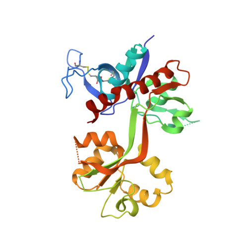
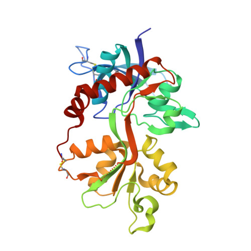
Structural Basis for Negative Allosteric Modulation of GluN2A-Containing NMDA Receptors.

Yi, F.Mou, T.C.Dorsett, K.N.Volkmann, R.A.Menniti, F.S.Sprang, S.R.Hansen, K.B.

(2016) Neuron 91: 1316-1329

  • DOI: https://doi.org/10.1016/j.neuron.2016.08.014
  • Primary Citation Related Structures: 
    5I56, 5I57, 5I58, 5I59, 5JTY

  • PubMed Abstract: 

    NMDA receptors mediate excitatory synaptic transmission and regulate synaptic plasticity in the central nervous system, but their dysregulation is also implicated in numerous brain disorders. Here, we describe GluN2A-selective negative allosteric modulators (NAMs) that inhibit NMDA receptors by stabilizing the apo state of the GluN1 ligand-binding domain (LBD), which is incapable of triggering channel gating. We describe structural determinants of NAM binding in crystal structures of the GluN1/2A LBD heterodimer, and analyses of NAM-bound LBD structures corresponding to active and inhibited receptor states reveal a molecular switch in the modulatory binding site that mediate the allosteric inhibition. NAM binding causes displacement of a valine in GluN2A and the resulting steric effects can be mitigated by the transition from glycine bound to apo state of the GluN1 LBD. This work provides mechanistic insight to allosteric NMDA receptor inhibition, thereby facilitating the development of novel classes NMDA receptor modulators as therapeutic agents.


  • Organizational Affiliation
    • Department of Biomedical and Pharmaceutical Sciences, University of Montana, Missoula, MT 59812, USA.

Macromolecule Content 

  • Total Structure Weight: 65.69 kDa 
  • Atom Count: 4,434 
  • Modeled Residue Count: 538 
  • Deposited Residue Count: 573 
  • Unique protein chains: 2

Macromolecules

Find similar proteins by:|  3D Structure
Entity ID: 1
MoleculeChains  Sequence LengthOrganismDetailsImage
Glutamate receptor ionotropic, NMDA 1,Glutamate receptor ionotropic, NMDA 1292Rattus norvegicusMutation(s): 0 
Gene Names: Grin1Nmdar1
UniProt
Find proteins for P35439 (Rattus norvegicus)
Explore P35439 
Go to UniProtKB:  P35439
Entity Groups
Sequence Clusters30% Identity50% Identity70% Identity90% Identity95% Identity100% Identity
UniProt GroupP35439
Sequence Annotations
Expand
Reference Sequence
Find similar proteins by:|  3D Structure
Entity ID: 2
MoleculeChains  Sequence LengthOrganismDetailsImage
Glutamate receptor ionotropic, NMDA 2A,Glutamate receptor ionotropic, NMDA 2A281Rattus norvegicusMutation(s): 0 
Gene Names: Grin2a
UniProt
Find proteins for Q00959 (Rattus norvegicus)
Explore Q00959 
Go to UniProtKB:  Q00959
Entity Groups
Sequence Clusters30% Identity50% Identity70% Identity90% Identity95% Identity100% Identity
UniProt GroupQ00959
Sequence Annotations
Expand
Reference Sequence

Small Molecules

Ligands 3 Unique
IDChains Name / Formula / InChI Key2D Diagram3D Interactions
6ND

Query on 6ND


Download:Ideal Coordinates CCD File
D [auth B]5-({[(3,4-difluorophenyl)sulfonyl]amino}methyl)-6-methyl-N-[(2-methyl-1,3-thiazol-5-yl)methyl]pyrazine-2-carboxamide
C18 H17 F2 N5 O3 S2
GZXSPNTWOSSNMG-UHFFFAOYSA-N
2JK

Query on 2JK


Download:Ideal Coordinates CCD File
C [auth A]4-hydroxy-5,7-dimethylquinoline-2-carboxylic acid
C12 H11 N O3
JLHAMAZZACUPTQ-UHFFFAOYSA-N
GLU

Query on GLU


Download:Ideal Coordinates CCD File
E [auth B]GLUTAMIC ACID
C5 H9 N O4
WHUUTDBJXJRKMK-VKHMYHEASA-N
Binding Affinity Annotations 
IDSourceBinding Affinity
6ND BindingDB:  5JTY IC50: 16.2 (nM) from 1 assay(s)

Experimental Data & Validation

Experimental Data

  • Method: X-RAY DIFFRACTION
  • Resolution: 2.72 Å
  • R-Value Free:  0.278 (Depositor), 0.274 (DCC) 
  • R-Value Work:  0.224 (Depositor), 0.225 (DCC) 
  • R-Value Observed: 0.230 (Depositor) 
Space Group: P 21 21 21
Unit Cell:
Length ( Å )Angle ( ˚ )
a = 54.569α = 90
b = 89.684β = 90
c = 130.415γ = 90
Software Package:
Software NamePurpose
PHENIXrefinement
HKL-2000data reduction
HKL-2000data scaling
PHASERphasing

Structure Validation

View Full Validation Report


Ligand Structure Quality Assessment 


Entry History 

Deposition Data

Revision History  (Full details and data files)

  • Version 1.0: 2016-09-21
    Type: Initial release
  • Version 1.1: 2016-10-05
    Changes: Database references
  • Version 1.2: 2023-09-27
    Changes: Data collection, Database references, Derived calculations, Refinement description
  • Version 1.3: 2024-10-23
    Changes: Structure summary