5JTI | pdb_00005jti

Crystal structure of the human Tankyrase 1 (TNKS) SAM domain (D1055R), crystal form 2


Experimental Data Snapshot

  • Method: X-RAY DIFFRACTION
  • Resolution: 2.90 Å
  • R-Value Free: 
    0.232 (Depositor), 0.196 (DCC) 
  • R-Value Work: 
    0.193 (Depositor), 0.203 (DCC) 
  • R-Value Observed: 
    0.195 (Depositor) 

Starting Model: experimental
View more details

wwPDB Validation 3D Report Full Report

Validation slider image for 5JTI

This is version 1.1 of the entry. See complete history

Literature

Tankyrase Requires SAM Domain-Dependent Polymerization to Support Wnt-beta-Catenin Signaling.

Mariotti, L.Templeton, C.M.Ranes, M.Paracuellos, P.Cronin, N.Beuron, F.Morris, E.Guettler, S.

(2016) Mol Cell 63: 498-513

  • DOI: https://doi.org/10.1016/j.molcel.2016.06.019
  • Primary Citation Related Structures: 
    5JRT, 5JTI, 5JU5

  • PubMed Abstract: 

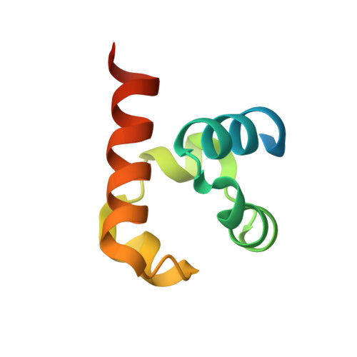
    The poly(ADP-ribose) polymerase (PARP) Tankyrase (TNKS and TNKS2) is paramount to Wnt-β-catenin signaling and a promising therapeutic target in Wnt-dependent cancers. The pool of active β-catenin is normally limited by destruction complexes, whose assembly depends on the polymeric master scaffolding protein AXIN. Tankyrase, which poly(ADP-ribosyl)ates and thereby destabilizes AXIN, also can polymerize, but the relevance of these polymers has remained unclear. We report crystal structures of the polymerizing TNKS and TNKS2 sterile alpha motif (SAM) domains, revealing versatile head-to-tail interactions. Biochemical studies informed by these structures demonstrate that polymerization is required for Tankyrase to drive β-catenin-dependent transcription. We show that the polymeric state supports PARP activity and allows Tankyrase to effectively access destruction complexes through enabling avidity-dependent AXIN binding. This study provides an example for regulated signal transduction in non-membrane-enclosed compartments (signalosomes), and it points to novel potential strategies to inhibit Tankyrase function in oncogenic Wnt signaling.


  • Organizational Affiliation
    • Division of Structural Biology, The Institute of Cancer Research (ICR), London SW7 3RP, UK; Division of Cancer Biology, The Institute of Cancer Research (ICR), London SW7 3RP, UK.

Macromolecule Content 

  • Total Structure Weight: 53.12 kDa 
  • Atom Count: 2,924 
  • Modeled Residue Count: 363 
  • Deposited Residue Count: 474 
  • Unique protein chains: 1

Macromolecules

Find similar proteins by:|  3D Structure
Entity ID: 1
MoleculeChains  Sequence LengthOrganismDetailsImage
Tankyrase-1
A, B, C, D, E
A, B, C, D, E, F
79Homo sapiensMutation(s): 1 
Gene Names: TNKSPARP5APARPLTIN1TINF1TNKS1
EC: 2.4.2.30 (PDB Primary Data), 2.4.2 (UniProt)
UniProt & NIH Common Fund Data Resources
Find proteins for O95271 (Homo sapiens)
Explore O95271 
Go to UniProtKB:  O95271
PHAROS:  O95271
GTEx:  ENSG00000173273 
Entity Groups
Sequence Clusters30% Identity50% Identity70% Identity90% Identity95% Identity100% Identity
UniProt GroupO95271
Sequence Annotations
Expand
Reference Sequence

Experimental Data & Validation

Experimental Data

  • Method: X-RAY DIFFRACTION
  • Resolution: 2.90 Å
  • R-Value Free:  0.232 (Depositor), 0.196 (DCC) 
  • R-Value Work:  0.193 (Depositor), 0.203 (DCC) 
  • R-Value Observed: 0.195 (Depositor) 
Space Group: P 1 21 1
Unit Cell:
Length ( Å )Angle ( ˚ )
a = 70.934α = 90
b = 55.48β = 102.9
c = 79.414γ = 90
Software Package:
Software NamePurpose
BUSTERrefinement
XDSdata reduction
Aimlessdata scaling
PHASERphasing

Structure Validation

View Full Validation Report


Entry History 

Deposition Data

Revision History  (Full details and data files)

  • Version 1.0: 2016-08-03
    Type: Initial release
  • Version 1.1: 2024-01-10
    Changes: Data collection, Database references, Refinement description