


Crystal structure of a Putative acyl-CoA dehydrogenase from Burkholderia xenovorans
Abendroth, J., Delker, S.L., Lorimer, D.D., Edewards, T.E.To be published.
Experimental Data Snapshot
Starting Model: experimental
View more details
Entity ID: 1 | |||||
|---|---|---|---|---|---|
| Molecule | Chains | Sequence Length | Organism | Details | Image |
| Putative acyl-CoA dehydrogenase | 410 | Paraburkholderia xenovorans LB400 | Mutation(s): 0 Gene Names: Bxe_B0278 EC: 1.3.8 | 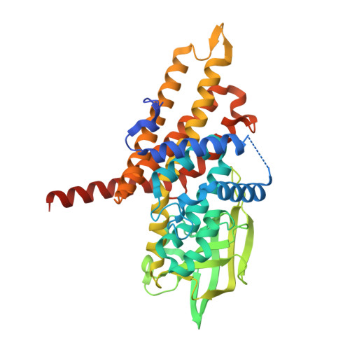 | |
UniProt | |||||
Entity Groups | |||||
| Sequence Clusters | 30% Identity50% Identity70% Identity90% Identity95% Identity100% Identity | ||||
| UniProt Group | Q13JS1 | ||||
Sequence AnnotationsExpand | |||||
Reference Sequence | |||||
| Ligands 4 Unique | |||||
|---|---|---|---|---|---|
| ID | Chains | Name / Formula / InChI Key | 2D Diagram | 3D Interactions | |
| FAD Download:Ideal Coordinates CCD File | E [auth A], H [auth B], M [auth C], T [auth D] | FLAVIN-ADENINE DINUCLEOTIDE C27 H33 N9 O15 P2 VWWQXMAJTJZDQX-UYBVJOGSSA-N |  | ||
| SO4 Download:Ideal Coordinates CCD File | K [auth B] L [auth B] P [auth C] Q [auth C] R [auth C] | SULFATE ION O4 S QAOWNCQODCNURD-UHFFFAOYSA-L |  | ||
| EDO Download:Ideal Coordinates CCD File | G [auth A], J [auth B], O [auth C], V [auth D] | 1,2-ETHANEDIOL C2 H6 O2 LYCAIKOWRPUZTN-UHFFFAOYSA-N |  | ||
| CL Download:Ideal Coordinates CCD File | F [auth A], I [auth B], N [auth C], U [auth D] | CHLORIDE ION Cl VEXZGXHMUGYJMC-UHFFFAOYSA-M |  | ||
| Length ( Å ) | Angle ( ˚ ) |
|---|---|
| a = 78.3 | α = 90 |
| b = 73.75 | β = 99.66 |
| c = 149.49 | γ = 90 |
| Software Name | Purpose |
|---|---|
| XDS | data reduction |
| XSCALE | data scaling |
| MOLREP | phasing |
| PHENIX | model building |
| Coot | model building |
| PHENIX | refinement |
| PDB_EXTRACT | data extraction |


