5JRB | pdb_00005jrb

Rad52(1-212) K102A/K133A/E202A mutant


Experimental Data Snapshot

  • Method: X-RAY DIFFRACTION
  • Resolution: 2.40 Å
  • R-Value Free: 
    0.273 (Depositor), 0.275 (DCC) 
  • R-Value Work: 
    0.224 (Depositor), 0.225 (DCC) 
  • R-Value Observed: 
    0.226 (Depositor) 

Starting Model: experimental
View more details

wwPDB Validation 3D Report Full Report

Validation slider image for 5JRB

This is version 1.2 of the entry. See complete history

Literature

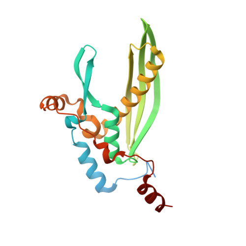
Structure of the human DNA-repair protein RAD52 containing surface mutations.

Saotome, M.Saito, K.Onodera, K.Kurumizaka, H.Kagawa, W.

(2016) Acta Crystallogr F Struct Biol Commun 72: 598-603

  • DOI: https://doi.org/10.1107/S2053230X1601027X
  • Primary Citation Related Structures: 
    5JRB

  • PubMed Abstract: 

    The Rad52 protein is a eukaryotic single-strand DNA-annealing protein that is involved in the homologous recombinational repair of DNA double-strand breaks. The isolated N-terminal half of the human RAD52 protein (RAD52(1-212)) forms an undecameric ring structure with a surface that is mostly positively charged. In the present study, it was found that RAD52(1-212) containing alanine mutations of the charged surface residues (Lys102, Lys133 and Glu202) is highly amenable to crystallization. The structure of the mutant RAD52(1-212) was solved at 2.4 Å resolution. The structure revealed an association between the symmetry-related RAD52(1-212) rings, in which a partially unfolded, C-terminal region of RAD52 extended into the DNA-binding groove of the neighbouring ring in the crystal. The alanine mutations probably reduced the surface entropy of the RAD52(1-212) ring and stabilized the ring-ring association observed in the crystal.


  • Organizational Affiliation
    • Department of Interdisciplinary Science and Engineering, Program in Chemistry and Life Science, School of Science and Engineering, Meisei University, 2-1-1 Hodokubo, Hino-shi, Tokyo 191-8506, Japan.

Macromolecule Content 

  • Total Structure Weight: 258.94 kDa 
  • Atom Count: 16,252 
  • Modeled Residue Count: 2,028 
  • Deposited Residue Count: 2,365 
  • Unique protein chains: 1

Macromolecules

Find similar proteins by:|  3D Structure
Entity ID: 1
MoleculeChains  Sequence LengthOrganismDetailsImage
DNA repair protein RAD52 homolog
A, B, C, D, E
A, B, C, D, E, F, G, H, I, J, K
215Homo sapiensMutation(s): 3 
Gene Names: RAD52
UniProt & NIH Common Fund Data Resources
Find proteins for P43351 (Homo sapiens)
Explore P43351 
Go to UniProtKB:  P43351
PHAROS:  P43351
GTEx:  ENSG00000002016 
Entity Groups
Sequence Clusters30% Identity50% Identity70% Identity90% Identity95% Identity100% Identity
UniProt GroupP43351
Sequence Annotations
Expand
Reference Sequence

Experimental Data & Validation

Experimental Data

  • Method: X-RAY DIFFRACTION
  • Resolution: 2.40 Å
  • R-Value Free:  0.273 (Depositor), 0.275 (DCC) 
  • R-Value Work:  0.224 (Depositor), 0.225 (DCC) 
  • R-Value Observed: 0.226 (Depositor) 
Space Group: P 1 21 1
Unit Cell:
Length ( Å )Angle ( ˚ )
a = 110.955α = 90
b = 98.516β = 91.99
c = 116.367γ = 90
Software Package:
Software NamePurpose
PHENIXrefinement
XDSdata reduction
MOLREPphasing

Structure Validation

View Full Validation Report


Entry History 

& Funding Information

Deposition Data


Funding OrganizationLocationGrant Number
JSPS KAKENHIJapan24570138
MEXT KAKENHIJapan26116521
MEXT KAKENHIJapan25116002

Revision History  (Full details and data files)

  • Version 1.0: 2016-08-10
    Type: Initial release
  • Version 1.1: 2019-12-25
    Changes: Data collection, Database references, Derived calculations
  • Version 1.2: 2023-11-08
    Changes: Data collection, Database references, Refinement description