De novo design of protein homo-oligomers with modular hydrogen-bond network-mediated specificity.
Boyken, S.E., Chen, Z., Groves, B., Langan, R.A., Oberdorfer, G., Ford, A., Gilmore, J.M., Xu, C., DiMaio, F., Pereira, J.H., Sankaran, B., Seelig, G., Zwart, P.H., Baker, D.(2016) Science 352: 680-687
- PubMed: 27151862 Search on PubMedSearch on PubMed Central
- DOI: https://doi.org/10.1126/science.aad8865
- Primary Citation Related Structures:
5IZS, 5J0H, 5J0I, 5J0J, 5J0K, 5J0L, 5J10, 5J2L, 5J73, 5JQZ - PubMed Abstract:
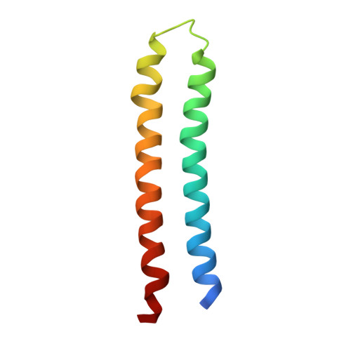
In nature, structural specificity in DNA and proteins is encoded differently: In DNA, specificity arises from modular hydrogen bonds in the core of the double helix, whereas in proteins, specificity arises largely from buried hydrophobic packing complemented by irregular peripheral polar interactions. Here, we describe a general approach for designing a wide range of protein homo-oligomers with specificity determined by modular arrays of central hydrogen-bond networks. We use the approach to design dimers, trimers, and tetramers consisting of two concentric rings of helices, including previously not seen triangular, square, and supercoiled topologies. X-ray crystallography confirms that the structures overall, and the hydrogen-bond networks in particular, are nearly identical to the design models, and the networks confer interaction specificity in vivo. The ability to design extensive hydrogen-bond networks with atomic accuracy enables the programming of protein interaction specificity for a broad range of synthetic biology applications; more generally, our results demonstrate that, even with the tremendous diversity observed in nature, there are fundamentally new modes of interaction to be discovered in proteins.
- Department of Biochemistry, University of Washington, Seattle, WA 98195, USA. Institute for Protein Design, University of Washington, Seattle, WA 98195, USA. Howard Hughes Medical Institute, University of Washington, Seattle, WA 98195, USA.
Organizational Affiliation: