5JPX | pdb_00005jpx

Solution structure of the TRIM21 B-box2 (B2)


Experimental Data Snapshot

  • Method: SOLUTION NMR
  • Conformers Calculated: 100 
  • Conformers Submitted: 20 
  • Selection Criteria: structures with the lowest energy 

wwPDB Validation 3D Report Full Report

Validation slider image for 5JPX

This is version 2.1 of the entry. See complete history

Literature

Solution NMR structure of the TRIM21 B-box2 and identification of residues involved in its interaction with the RING domain.

Wallenhammar, A.Anandapadamanaban, M.Lemak, A.Mirabello, C.Lundstrom, P.Wallner, B.Sunnerhagen, M.

(2017) PLoS One 12: e0181551-e0181551

  • DOI: https://doi.org/10.1371/journal.pone.0181551
  • Primary Citation Related Structures: 
    5JPX

  • PubMed Abstract: 

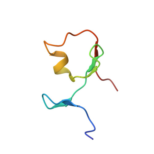
    Tripartite motif-containing (TRIM) proteins are defined by the sequential arrangement of RING, B-box and coiled-coil domains (RBCC), where the B-box domain is a unique feature of the TRIM protein family. TRIM21 is an E3 ubiquitin-protein ligase implicated in innate immune signaling by acting as an autoantigen and by modifying interferon regulatory factors. Here we report the three-dimensional solution structure of the TRIM21 B-box2 domain by nuclear magnetic resonance (NMR) spectroscopy. The structure of the B-box2 domain, comprising TRIM21 residues 86-130, consists of a short α-helical segment with an N-terminal short β-strand and two anti-parallel β-strands jointly found the core, and adopts a RING-like fold. This ββαβ core largely defines the overall fold of the TRIM21 B-box2 and the coordination of one Zn2+ ion stabilizes the tertiary structure of the protein. Using NMR titration experiments, we have identified an exposed interaction surface, a novel interaction patch where the B-box2 is likely to bind the N-terminal RING domain. Our structure together with comparisons with other TRIM B-box domains jointly reveal how its different surfaces are employed for various modular interactions, and provides extended understanding of how this domain relates to flanking domains in TRIM proteins.


  • Organizational Affiliation
    • Division of Chemistry, Department of Physics, Chemistry and Biology, Linköping University, Linköping, Sweden.

Macromolecule Content 

  • Total Structure Weight: 5.21 kDa 
  • Atom Count: 356 
  • Modeled Residue Count: 45 
  • Deposited Residue Count: 45 
  • Unique protein chains: 1

Macromolecules

Find similar proteins by:|  3D Structure
Entity ID: 1
MoleculeChains  Sequence LengthOrganismDetailsImage
E3 ubiquitin-protein ligase TRIM2145Homo sapiensMutation(s): 0 
Gene Names: TRIM21RNF81RO52SSA1
EC: 6.3.2 (PDB Primary Data), 2.3.2.27 (UniProt)
UniProt & NIH Common Fund Data Resources
Find proteins for P19474 (Homo sapiens)
Explore P19474 
Go to UniProtKB:  P19474
PHAROS:  P19474
GTEx:  ENSG00000132109 
Entity Groups
Sequence Clusters30% Identity50% Identity70% Identity90% Identity95% Identity100% Identity
UniProt GroupP19474
Sequence Annotations
Expand
Reference Sequence

Small Molecules

Ligands 1 Unique
IDChains Name / Formula / InChI Key2D Diagram3D Interactions
ZN

Query on ZN


Download:Ideal Coordinates CCD File
B [auth A]ZINC ION
Zn
PTFCDOFLOPIGGS-UHFFFAOYSA-N

Experimental Data & Validation

Experimental Data

  • Method: SOLUTION NMR
  • Conformers Calculated: 100 
  • Conformers Submitted: 20 
  • Selection Criteria: structures with the lowest energy 

Structure Validation

View Full Validation Report


Entry History 

& Funding Information

Deposition Data


Funding OrganizationLocationGrant Number
Swedish Research CouncilSweden--
CancerfondenSweden--

Revision History  (Full details and data files)

  • Version 1.0: 2017-08-09
    Type: Initial release
  • Version 1.1: 2017-09-13
    Changes: Author supporting evidence
  • Version 1.2: 2018-02-07
    Changes: Experimental preparation
  • Version 1.3: 2019-05-08
    Changes: Data collection
  • Version 2.0: 2019-10-30
    Changes: Atomic model, Data collection, Database references
  • Version 2.1: 2024-06-19
    Changes: Data collection, Database references