5JNB | pdb_00005jnb

structure of GLD-2/RNP-8 complex


Experimental Data Snapshot

  • Method: X-RAY DIFFRACTION
  • Resolution: 2.49 Å
  • R-Value Free: 
    0.242 (Depositor), 0.244 (DCC) 
  • R-Value Work: 
    0.192 (Depositor), 0.198 (DCC) 
  • R-Value Observed: 
    0.194 (Depositor) 

Starting Model: experimental
View more details

wwPDB Validation 3D Report Full Report

Validation slider image for 5JNB

This is version 1.4 of the entry. See complete history

Literature

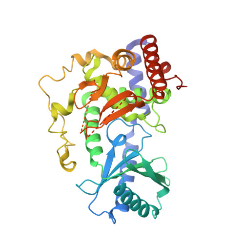
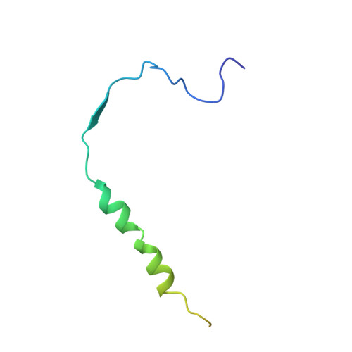
Structural basis for the antagonistic roles of RNP-8 and GLD-3 in GLD-2 poly(A)-polymerase activity.

Nakel, K.Bonneau, F.Basquin, C.Habermann, B.Eckmann, C.R.Conti, E.

(2016) RNA 22: 1139-1145

  • DOI: https://doi.org/10.1261/rna.056598.116
  • Primary Citation Related Structures: 
    5JNB

  • PubMed Abstract: 

    Cytoplasmic polyadenylation drives the translational activation of specific mRNAs in early metazoan development and is performed by distinct complexes that share the same catalytic poly(A)-polymerase subunit, GLD-2. The activity and specificity of GLD-2 depend on its binding partners. In Caenorhabditis elegans, GLD-2 promotes spermatogenesis when bound to GLD-3 and oogenesis when bound to RNP-8. GLD-3 and RNP-8 antagonize each other and compete for GLD-2 binding. Following up on our previous mechanistic studies of GLD-2-GLD-3, we report here the 2.5 Å resolution structure and biochemical characterization of a GLD-2-RNP-8 core complex. In the structure, RNP-8 embraces the poly(A)-polymerase, docking onto several conserved hydrophobic hotspots present on the GLD-2 surface. RNP-8 stabilizes GLD-2 and indirectly stimulates polyadenylation. RNP-8 has a different amino-acid sequence and structure as compared to GLD-3. Yet, it binds the same surfaces of GLD-2 by forming alternative interactions, rationalizing the remarkable versatility of GLD-2 complexes.


  • Organizational Affiliation
    • Department of Structural Cell Biology, Max-Planck-Institute of Biochemistry, D-82152 Martinsried, Germany.

Macromolecule Content 

  • Total Structure Weight: 189.73 kDa 
  • Atom Count: 10,776 
  • Modeled Residue Count: 1,350 
  • Deposited Residue Count: 1,648 
  • Unique protein chains: 2

Macromolecules

Find similar proteins by:|  3D Structure
Entity ID: 1
MoleculeChains  Sequence LengthOrganismDetailsImage
Poly(A) RNA polymerase gld-2
A, B, C, D
338Caenorhabditis elegansMutation(s): 1 
Gene Names: gld-2ZC308.1
EC: 2.7.7.19
UniProt
Find proteins for O17087 (Caenorhabditis elegans)
Explore O17087 
Go to UniProtKB:  O17087
Entity Groups
Sequence Clusters30% Identity50% Identity70% Identity90% Identity95% Identity100% Identity
UniProt GroupO17087
Sequence Annotations
Expand
Reference Sequence
Find similar proteins by:|  3D Structure
Entity ID: 2
MoleculeChains  Sequence LengthOrganismDetailsImage
RNP (RRM RNA binding domain) containing
E, F, G, H
74Caenorhabditis elegansMutation(s): 0 
Gene Names: rnp-8CELE_R119.7R119.7
UniProt
Find proteins for O61711 (Caenorhabditis elegans)
Explore O61711 
Go to UniProtKB:  O61711
Entity Groups
Sequence Clusters30% Identity50% Identity70% Identity90% Identity95% Identity100% Identity
UniProt GroupO61711
Sequence Annotations
Expand
Reference Sequence

Small Molecules

Ligands 3 Unique
IDChains Name / Formula / InChI Key2D Diagram3D Interactions
SO4

Query on SO4


Download:Ideal Coordinates CCD File
I [auth A],
N [auth B],
S [auth C],
V [auth D]
SULFATE ION
O4 S
QAOWNCQODCNURD-UHFFFAOYSA-L
EDO

Query on EDO


Download:Ideal Coordinates CCD File
K [auth A]
L [auth A]
M [auth A]
O [auth B]
P [auth B]
K [auth A],
L [auth A],
M [auth A],
O [auth B],
P [auth B],
Q [auth B],
T [auth C],
U [auth C],
W [auth D]
1,2-ETHANEDIOL
C2 H6 O2
LYCAIKOWRPUZTN-UHFFFAOYSA-N
MG

Query on MG


Download:Ideal Coordinates CCD File
J [auth A],
R [auth B]
MAGNESIUM ION
Mg
JLVVSXFLKOJNIY-UHFFFAOYSA-N

Experimental Data & Validation

Experimental Data

  • Method: X-RAY DIFFRACTION
  • Resolution: 2.49 Å
  • R-Value Free:  0.242 (Depositor), 0.244 (DCC) 
  • R-Value Work:  0.192 (Depositor), 0.198 (DCC) 
  • R-Value Observed: 0.194 (Depositor) 
Space Group: P 1
Unit Cell:
Length ( Å )Angle ( ˚ )
a = 65.347α = 89.88
b = 86.435β = 93.13
c = 91.907γ = 90.22
Software Package:
Software NamePurpose
PHENIXrefinement
XDSdata reduction
XDSdata scaling
PHENIXphasing

Structure Validation

View Full Validation Report


Entry History 

& Funding Information

Deposition Data


Funding OrganizationLocationGrant Number
German Research FoundationGermanySFB646
German Research FoundationGermanySFB1035
German Research FoundationGermanyGRK1721
German Research FoundationGermanyFOR1680
European CommissionGermanyERC Advanced Investigator Grant 294371
European CommissionGermanyMarie Curie ITN RNPnet
German Research FoundationGermanyEC369/3-1
German Research FoundationGermanyGRK1591
German Research FoundationGermanyBU2451/1-2

Revision History  (Full details and data files)

  • Version 1.0: 2016-06-22
    Type: Initial release
  • Version 1.1: 2016-07-06
    Changes: Data collection
  • Version 1.2: 2016-07-27
    Changes: Database references
  • Version 1.3: 2017-09-06
    Changes: Author supporting evidence
  • Version 1.4: 2024-01-10
    Changes: Data collection, Database references, Refinement description