5JE8

The crystal structure of Bacillus cereus 3-hydroxyisobutyrate dehydrogenase in complex with NAD


Experimental Data Snapshot

  • Method: X-RAY DIFFRACTION
  • Resolution: 2.10 Å
  • R-Value Free: 0.243 
  • R-Value Work: 0.197 
  • R-Value Observed: 0.200 

Starting Model: experimental
View more details

wwPDB Validation   3D Report Full Report


Ligand Structure Quality Assessment 


This is version 1.2 of the entry. See complete history


Literature

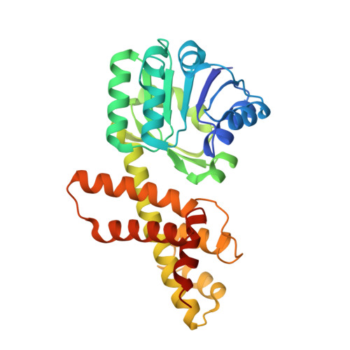
Structural and biochemical characterization of the Bacillus cereus 3-hydroxyisobutyrate dehydrogenase

Park, S.C.Kim, P.H.Lee, G.S.Kang, S.G.Ko, H.J.Yoon, S.I.

(2016) Biochem Biophys Res Commun 474: 522-527

  • DOI: https://doi.org/10.1016/j.bbrc.2016.04.126
  • Primary Citation of Related Structures:  
    5JE8

  • PubMed Abstract: 

    The 3-hydroxyisobutyrate dehydrogenase (HIBADH) family catalyzes the NAD(+)- or NADP(+)-dependent oxidation of various β-hydroxyacid substrates into their cognate semialdehydes for diverse metabolic pathways. Because HIBADH group members exhibit different substrate specificities, the substrate-recognition mode of each enzyme should be individually characterized. In the current study, we report the biochemical and structural analysis of a HIBADH group enzyme from Bacillus cereus (bcHIBADH). bcHIBADH mediates a dehydrogenation reaction on S-3-hydroxyisobutyrate substrate with high catalytic efficiency in an NAD(+)-dependent manner; it also oxidizes l-serine and 3-hydroxypropionate with lower activity. bcHIBADH consists of two domains and is further assembled into a functional dimer rather than a tetramer that has been commonly observed in other prokaryotic HIBADH group members. In the bcHIBADH structure, the interdomain cleft forms a putative active site and simultaneously accommodates both an NAD(+) cofactor and a substrate mimic. Our structure-based comparative analysis highlights structural motifs that are important in the cofactor and substrate recognition of the HIBADH group.


  • Organizational Affiliation

    Division of Biomedical Convergence, College of Biomedical Science, Kangwon National University, Chuncheon 24341, Republic of Korea.


Macromolecules
Find similar proteins by:  (by identity cutoff)  |  3D Structure
Entity ID: 1
MoleculeChains Sequence LengthOrganismDetailsImage
3-hydroxyisobutyrate dehydrogenase
A, B, C, D
302Bacillus cereus ATCC 14579Mutation(s): 0 
Gene Names: BC_2289
EC: 1.1.1.31
UniProt
Find proteins for Q81DR6 (Bacillus cereus (strain ATCC 14579 / DSM 31 / CCUG 7414 / JCM 2152 / NBRC 15305 / NCIMB 9373 / NCTC 2599 / NRRL B-3711))
Explore Q81DR6 
Go to UniProtKB:  Q81DR6
Entity Groups  
Sequence Clusters30% Identity50% Identity70% Identity90% Identity95% Identity100% Identity
UniProt GroupQ81DR6
Sequence Annotations
Expand
  • Reference Sequence
Experimental Data & Validation

Experimental Data

  • Method: X-RAY DIFFRACTION
  • Resolution: 2.10 Å
  • R-Value Free: 0.243 
  • R-Value Work: 0.197 
  • R-Value Observed: 0.200 
  • Space Group: P 21 21 21
Unit Cell:
Length ( Å )Angle ( ˚ )
a = 46.444α = 90
b = 158.576β = 90
c = 163.138γ = 90
Software Package:
Software NamePurpose
REFMACrefinement
HKL-2000data reduction
HKL-2000data scaling
PHASERphasing

Structure Validation

View Full Validation Report


Ligand Structure Quality Assessment 


Entry History 

Deposition Data

Revision History  (Full details and data files)

  • Version 1.0: 2016-05-11
    Type: Initial release
  • Version 1.1: 2016-06-01
    Changes: Database references
  • Version 1.2: 2023-11-08
    Changes: Data collection, Database references, Derived calculations, Refinement description