5JD2 | pdb_00005jd2

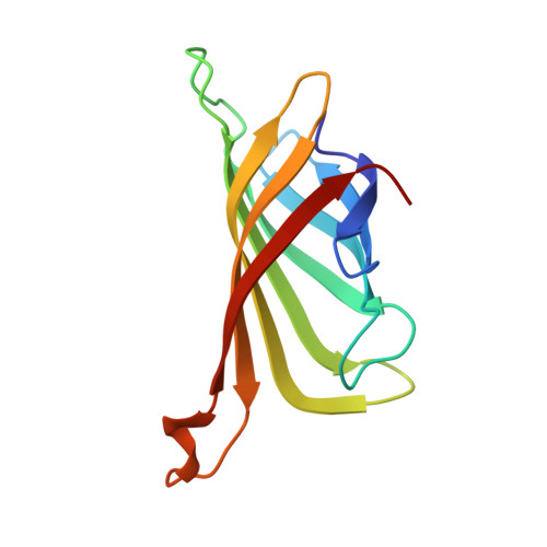
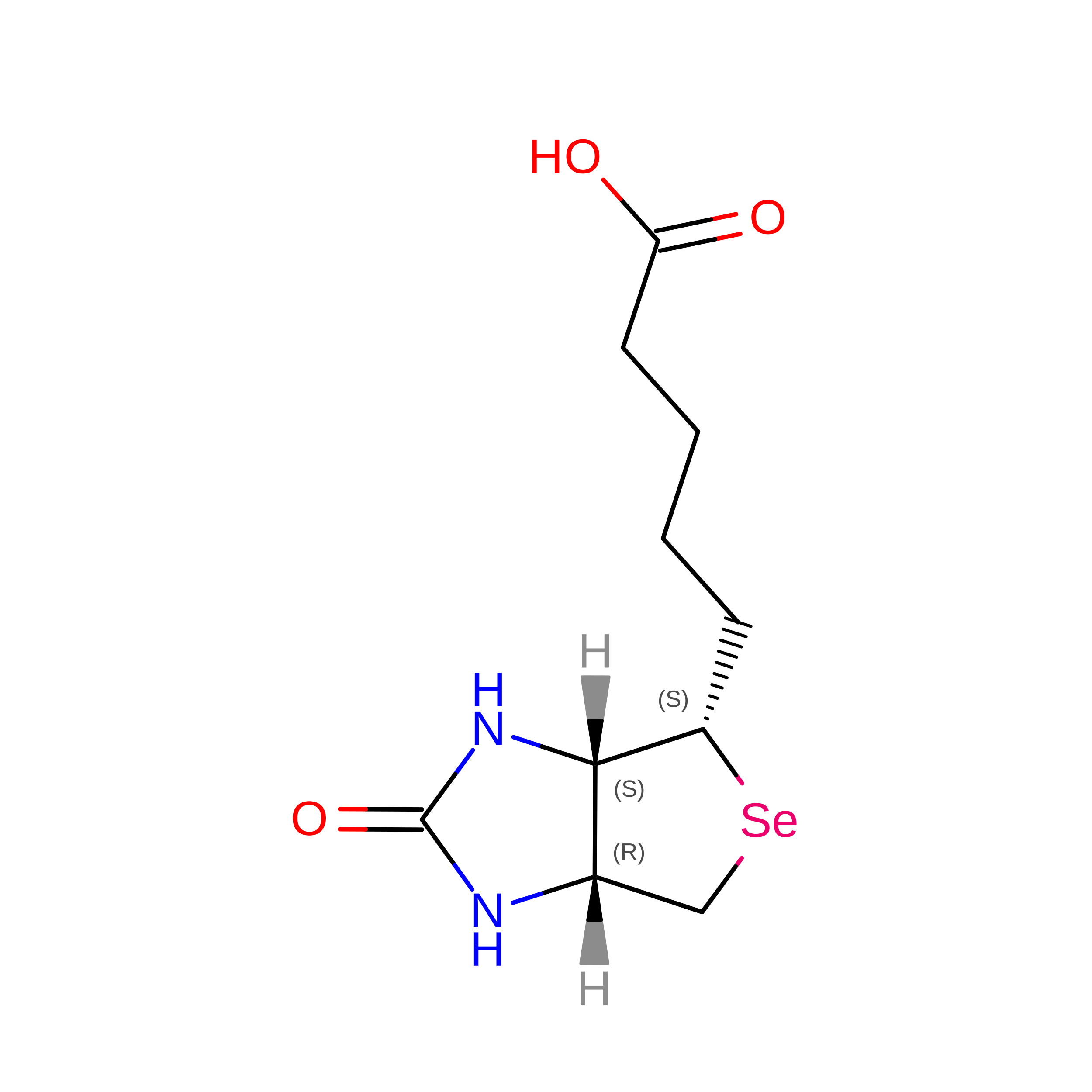
SFX structure of corestreptavidin-selenobiotin complex


Experimental Data Snapshot

  • Method: X-RAY DIFFRACTION
  • Resolution: 1.90 Å
  • R-Value Free: 
    0.200 (Depositor), 0.204 (DCC) 
  • R-Value Work: 
    0.165 (Depositor), 0.167 (DCC) 
  • R-Value Observed: 
    0.166 (Depositor) 

wwPDB Validation 3D Report Full Report

Validation slider image for 5JD2

Ligand Structure Quality Assessment 


This is version 1.5 of the entry. See complete history. 

Literature

Selenium single-wavelength anomalous diffraction de novo phasing using an X-ray-free electron laser.

Hunter, M.S., Yoon, C.H., DeMirci, H., Sierra, R.G., Dao, E.H., Ahmadi, R., Aksit, F., Aquila, A.L., Ciftci, H., Guillet, S., Hayes, M.J., Lane, T.J., Liang, M., Lundstrom, U., Koglin, J.E., Mgbam, P., Rao, Y., Zhang, L., Wakatsuki, S., Holton, J.M., Boutet, S.

(2016) Nat Commun 7: 13388-13388

  • DOI: https://doi.org/10.1038/ncomms13388
  • Primary Citation Related Structures: 
    5JD2

  • PubMed Abstract: 

    Structural information about biological macromolecules near the atomic scale provides important insight into the functions of these molecules. To date, X-ray crystallography has been the predominant method used for macromolecular structure determination. However, challenges exist when solving structures with X-rays, including the phase problem and radiation damage. X-ray-free electron lasers (X-ray FELs) have enabled collection of diffraction information before the onset of radiation damage, yet the majority of structures solved at X-ray FELs have been phased using external information via molecular replacement. De novo phasing at X-ray FELs has proven challenging due in part to per-pulse variations in intensity and wavelength. Here we report the solution of a selenobiotinyl-streptavidin structure using phases obtained by the anomalous diffraction of selenium measured at a single wavelength (Se-SAD) at the Linac Coherent Light Source. Our results demonstrate Se-SAD, routinely employed at synchrotrons for novel structure determination, is now possible at X-ray FELs.


  • Organizational Affiliation: 
    • Linac Coherent Light Source, SLAC National Accelerator Laboratory, Menlo Park, California 94025, USA.

Macromolecule Content 

  • Total Structure Weight: 51.94 kDa 
  • Atom Count: 3,959 
  • Modeled Residue Count: 474 
  • Deposited Residue Count: 480 
  • Unique protein chains: 1

Macromolecules

Find similar proteins by:|  3D Structure
Entity ID: 1
MoleculeChains  Sequence LengthOrganismDetailsImage
Streptavidin
A, B, C, D
120Streptomyces avidiniiMutation(s): 0 
UniProt
Find proteins for P22629 (Streptomyces avidinii)
Explore P22629 
Go to UniProtKB:  P22629
Entity Groups
Sequence Clusters30% Identity50% Identity70% Identity90% Identity95% Identity100% Identity
UniProt GroupP22629
Sequence Annotations
Expand
Reference Sequence

Experimental Data & Validation

Experimental Data

  • Method: X-RAY DIFFRACTION
  • Resolution: 1.90 Å
  • R-Value Free:  0.200 (Depositor), 0.204 (DCC) 
  • R-Value Work:  0.165 (Depositor), 0.167 (DCC) 
  • R-Value Observed: 0.166 (Depositor) 
Space Group: P 1 21 1
Unit Cell:
Length ( Å )Angle ( ˚ )
a = 50.84α = 90
b = 98.52β = 112.38
c = 53.43γ = 90
Software Package:
Software NamePurpose
DMphasing
PHENIXrefinement
PDB_EXTRACTdata extraction
CrystFELdata reduction
CrystFELdata scaling

Structure Validation

View Full Validation Report


Ligand Structure Quality Assessment 


Entry History 

& Funding Information

Deposition Data


Funding OrganizationLocationGrant Number
Department of Energy (DOE, United States)United States--

Revision History  (Full details and data files)

  • Version 1.0: 2016-11-16
    Type: Initial release
  • Version 1.1: 2017-09-06
    Changes: Advisory, Author supporting evidence, Derived calculations
  • Version 1.2: 2017-11-22
    Changes: Refinement description
  • Version 1.3: 2018-02-14
    Changes: Data collection
  • Version 1.4: 2019-12-04
    Changes: Author supporting evidence
  • Version 1.5: 2024-03-06
    Changes: Advisory, Data collection, Database references