5J9A

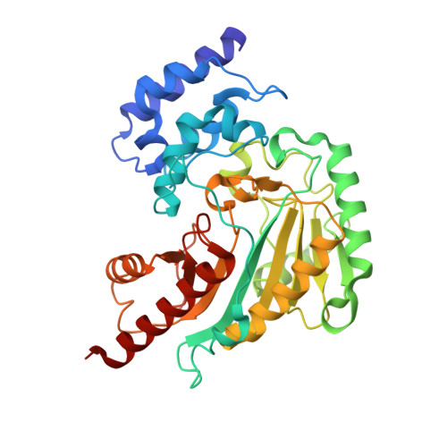
Ambient temperature transition state structure of arginine kinase - crystal 11/Form II


Experimental Data Snapshot

  • Method: X-RAY DIFFRACTION
  • Resolution: 2.00 Å
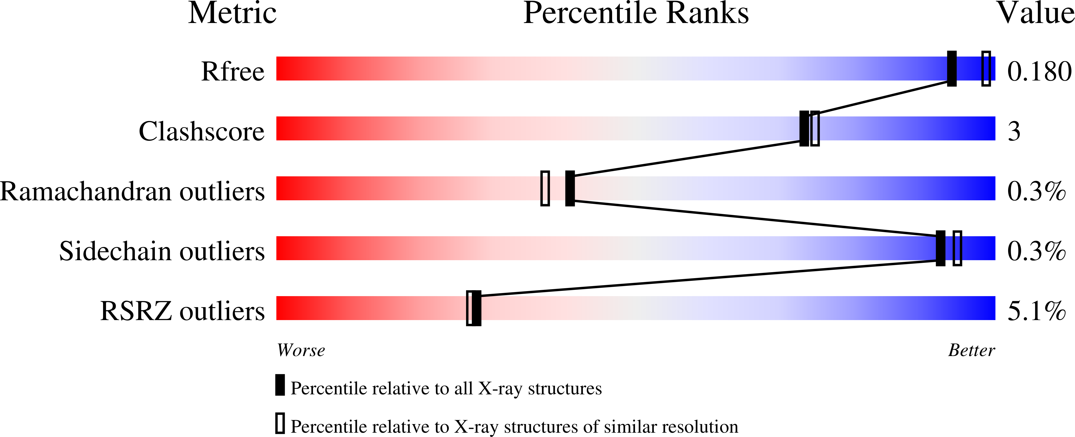
  • R-Value Free: 0.179 
  • R-Value Work: 0.135 
  • R-Value Observed: 0.138 

Starting Model: experimental
View more details

wwPDB Validation   3D Report Full Report


Ligand Structure Quality Assessment 


This is version 1.5 of the entry. See complete history


Literature

The Sampling of Conformational Dynamics in Ambient-Temperature Crystal Structures of Arginine Kinase.

Godsey, M.H.Davulcu, O.Nix, J.C.Skalicky, J.J.Bruschweiler, R.P.Chapman, M.S.

(2016) Structure 24: 1658-1667

  • DOI: https://doi.org/10.1016/j.str.2016.07.013
  • Primary Citation of Related Structures:  
    5J99, 5J9A

  • PubMed Abstract: 

    Arginine kinase provides a model for functional dynamics, studied through crystallography, enzymology, and nuclear magnetic resonance. Structures are now solved, at ambient temperature, for the transition state analog (TSA) complex. Analysis of quasi-rigid sub-domain displacements show that differences between the two TSA structures average about 5% of changes between substrate-free and TSA forms, and they are nearly co-linear. Small backbone hinge rotations map to sites that also flex on substrate binding. Anisotropic atomic displacement parameters (ADPs) are refined using rigid-body TLS constraints. Consistency between crystal forms shows that they reflect intrinsic molecular properties more than crystal lattice effects. In many regions, the favored directions of thermal/static displacement are appreciably correlated with movements on substrate binding. Correlation between ADPs and larger substrate-associated movements implies that the latter approximately follow paths of low-energy intrinsic motions.


  • Organizational Affiliation

    Department of Math/Science, Concordia University, Portland, OR 97211, USA.


Macromolecules
Find similar proteins by:  (by identity cutoff)  |  3D Structure
Entity ID: 1
MoleculeChains Sequence LengthOrganismDetailsImage
Arginine kinase357Limulus polyphemusMutation(s): 3 
EC: 2.7.3.3
UniProt
Find proteins for P51541 (Limulus polyphemus)
Explore P51541 
Go to UniProtKB:  P51541
Entity Groups  
Sequence Clusters30% Identity50% Identity70% Identity90% Identity95% Identity100% Identity
UniProt GroupP51541
Sequence Annotations
Expand
  • Reference Sequence
Experimental Data & Validation

Experimental Data

  • Method: X-RAY DIFFRACTION
  • Resolution: 2.00 Å
  • R-Value Free: 0.179 
  • R-Value Work: 0.135 
  • R-Value Observed: 0.138 
  • Space Group: P 21 21 21
Unit Cell:
Length ( Å )Angle ( ˚ )
a = 66.038α = 90
b = 72.047β = 90
c = 80.237γ = 90
Software Package:
Software NamePurpose
PHENIXrefinement
XDSdata reduction
XDSdata scaling
PHASERphasing

Structure Validation

View Full Validation Report


Ligand Structure Quality Assessment 


Entry History & Funding Information

Deposition Data


Funding OrganizationLocationGrant Number
National Institutes of Health/National Institute of General Medical Sciences (NIH/NIGMS)United StatesGM77643

Revision History  (Full details and data files)

  • Version 1.0: 2016-08-17
    Type: Initial release
  • Version 1.1: 2016-09-21
    Changes: Database references
  • Version 1.2: 2016-10-19
    Changes: Database references
  • Version 1.3: 2017-09-13
    Changes: Author supporting evidence, Database references, Derived calculations
  • Version 1.4: 2019-12-25
    Changes: Author supporting evidence
  • Version 1.5: 2023-09-27
    Changes: Data collection, Database references, Derived calculations, Refinement description