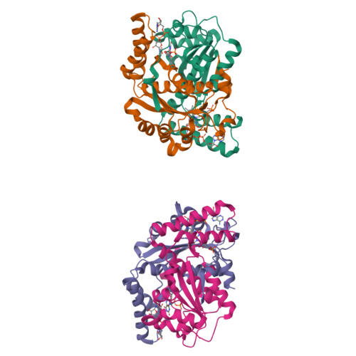
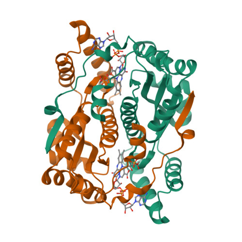
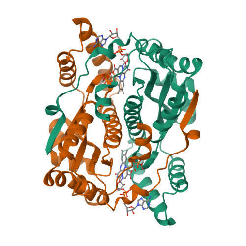
5J8D

Structure of nitroreductase from E. cloacae complexed with nicotinic acid adenine dinucleotide


Experimental Data Snapshot

  • Method: X-RAY DIFFRACTION
  • Resolution: 1.85 Å
  • R-Value Free: 0.212 
  • R-Value Work: 0.175 
  • R-Value Observed: 0.178 

Starting Model: experimental
View more details

wwPDB Validation   3D Report Full Report


Ligand Structure Quality Assessment 


This is version 1.4 of the entry. See complete history


Literature

Mechanism-Informed Refinement Reveals Altered Substrate-Binding Mode for Catalytically Competent Nitroreductase.

Pitsawong, W.Haynes, C.A.Koder, R.L.Rodgers, D.W.Miller, A.F.

(2017) Structure 25: 978-987.e4

  • DOI: https://doi.org/10.1016/j.str.2017.05.002
  • Primary Citation of Related Structures:  
    5J8D, 5J8G

  • PubMed Abstract: 

    Nitroreductase (NR) from Enterobacter cloacae reduces diverse nitroaromatics including herbicides, explosives, and prodrugs, and holds promise for bioremediation, prodrug activation, and enzyme-assisted synthesis. We solved crystal structures of NR complexes with bound substrate or analog for each of its two half-reactions. We complemented these with kinetic isotope effect (KIE) measurements elucidating H-transfer steps essential to each half-reaction. KIEs indicate hydride transfer from NADH to the flavin consistent with our structure of NR with the NADH analog nicotinic acid adenine dinucleotide (NAAD). The KIE on reduction of p-nitrobenzoic acid (p-NBA) also indicates hydride transfer, and requires revision of prior computational mechanisms. Our mechanistic information provided a structural restraint for the orientation of bound substrate, placing the nitro group closer to the flavin N5 in the pocket that binds the amide of NADH. KIEs show that solvent provides a proton, enabling accommodation of different nitro group placements, consistent with the broad repertoire of NR.


  • Organizational Affiliation

    Department of Chemistry, University of Kentucky, 505 Rose Street, Lexington, KY 40506-0055, USA.


Macromolecules
Find similar proteins by:  (by identity cutoff)  |  3D Structure
Entity ID: 1
MoleculeChains Sequence LengthOrganismDetailsImage
Oxygen-insensitive NAD(P)H nitroreductase
A, B, C, D
216Enterobacter cloacaeMutation(s): 0 
Gene Names: nfsBnfnBnfsI
EC: 1
UniProt
Find proteins for Q01234 (Enterobacter cloacae)
Explore Q01234 
Go to UniProtKB:  Q01234
Entity Groups  
Sequence Clusters30% Identity50% Identity70% Identity90% Identity95% Identity100% Identity
UniProt GroupQ01234
Sequence Annotations
Expand
  • Reference Sequence
Experimental Data & Validation

Experimental Data

  • Method: X-RAY DIFFRACTION
  • Resolution: 1.85 Å
  • R-Value Free: 0.212 
  • R-Value Work: 0.175 
  • R-Value Observed: 0.178 
  • Space Group: P 1 21 1
Unit Cell:
Length ( Å )Angle ( ˚ )
a = 49.38α = 90
b = 113.14β = 92.59
c = 80.7γ = 90
Software Package:
Software NamePurpose
PHENIXrefinement
PDB_EXTRACTdata extraction
HKL-2000data reduction
HKL-2000data scaling
CNSphasing

Structure Validation

View Full Validation Report


Ligand Structure Quality Assessment 


Entry History & Funding Information

Deposition Data


Funding OrganizationLocationGrant Number
National Institutes of Health/National Institute of Neurological Disorders and Stroke (NIH/NINDS)United StatesNS38401
National Science Foundation (NSF, United States)United StatesMCB 9904886
National Institutes of Health/National Institute of General Medical Sciences (NIH/NIGMS)United StatesGM110787
National Science Foundation (NSF, United States)United StatesIIA-1355438

Revision History  (Full details and data files)

  • Version 1.0: 2017-05-17
    Type: Initial release
  • Version 1.1: 2017-09-20
    Changes: Author supporting evidence
  • Version 1.2: 2017-12-06
    Changes: Database references
  • Version 1.3: 2019-11-27
    Changes: Author supporting evidence
  • Version 1.4: 2023-09-27
    Changes: Data collection, Database references, Refinement description