Development of Dihydroxyphenyl Sulfonylisoindoline Derivatives as Liver-Targeting Pyruvate Dehydrogenase Kinase Inhibitors.
Tso, S.C., Lou, M., Wu, C.Y., Gui, W.J., Chuang, J.L., Morlock, L.K., Williams, N.S., Wynn, R.M., Qi, X., Chuang, D.T.(2017) J Med Chem 60: 1142-1150
- PubMed: 28085286 Search on PubMedSearch on PubMed Central
- DOI: https://doi.org/10.1021/acs.jmedchem.6b01540
- Primary Citation Related Structures:
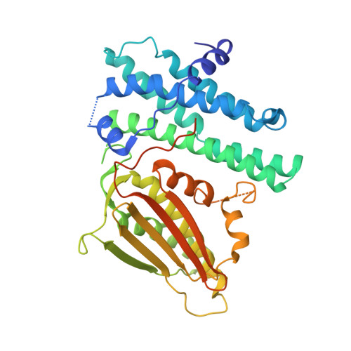
5J6A, 5J71 - PubMed Abstract:
Pyruvate dehydrogenase kinases 1-4 (PDK1-4) negatively control activity of the pyruvate dehydrogenase complex (PDC) and are up-regulated in obesity, diabetes, heart failure, and cancer. We reported earlier two novel pan-PDK inhibitors PS8 [4-((5-hydroxyisoindolin-2-yl)sulfonyl)benzene-1,3-diol] (1) and PS10 [2-((2,4-dihydroxyphenyl)sulfonyl)isoindoline-4,6-diol] (2) that targeted the ATP-binding pocket in PDKs. Here, we developed a new generation of PDK inhibitors by extending the dihydroxyphenyl sulfonylisoindoline scaffold in 1 and 2 to the entrance region of the ATP-binding pocket in PDK2. The lead inhibitor (S)-3-amino-4-(4-((2-((2,4-dihydroxyphenyl)sulfonyl)isoindolin-5-yl)amino)piperidin-1-yl)-4-oxobutanamide (17) shows a ∼8-fold lower IC 50 (58 nM) than 2 (456 nM). In the crystal structure, the asparagine moiety in 17 provides additional interactions with Glu-262 from PDK2. Treatment of diet-induced obese mice with 17 resulted in significant liver-specific augmentation of PDC activity, accompanied by improved glucose tolerance and drastically reduced hepatic steatosis. These findings support 17 as a potential glucose-lowering therapeutic targeting liver for obesity and type 2 diabetes.
- Chemistry Center, National Institute of Biological Science , Beijing 102206, China.
Organizational Affiliation: