5J5T

GLK co-crystal structure with aminopyrrolopyrimidine inhibitor


Experimental Data Snapshot

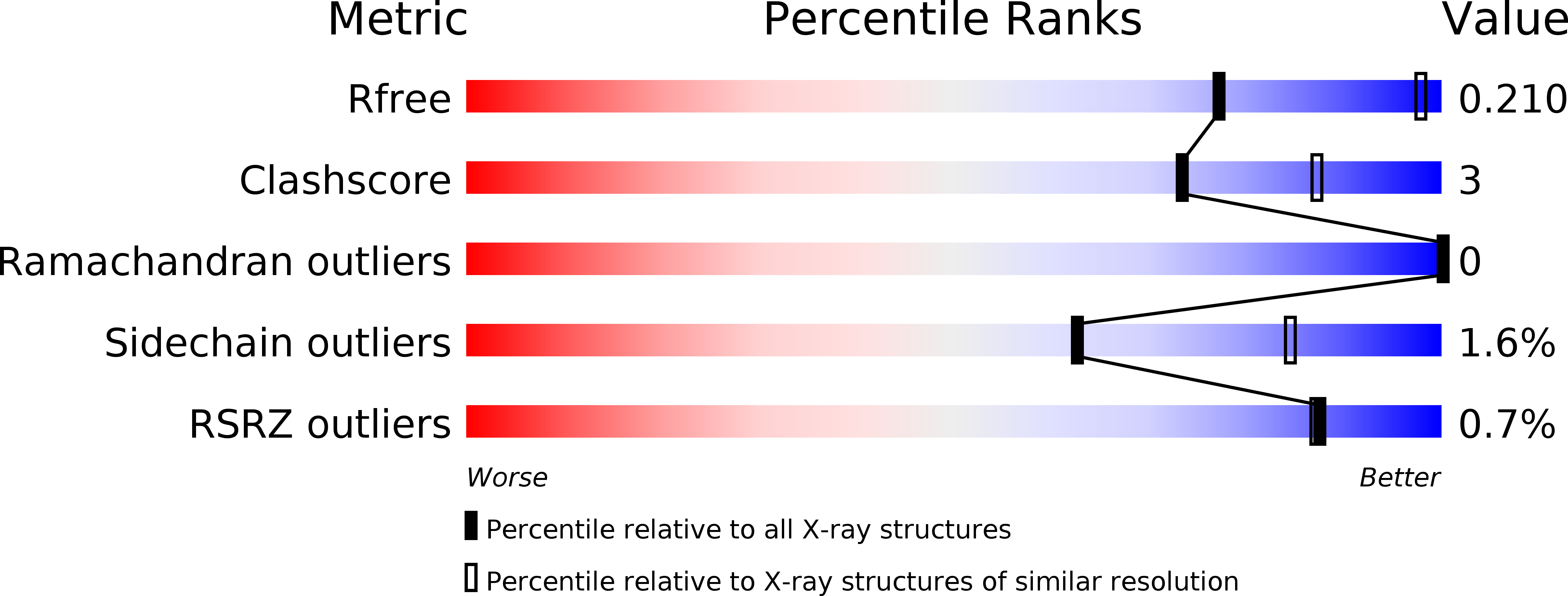
  • Method: X-RAY DIFFRACTION
  • Resolution: 2.85 Å
  • R-Value Free: 0.206 
  • R-Value Work: 0.175 
  • R-Value Observed: 0.177 

Starting Model: experimental
View more details

wwPDB Validation   3D Report Full Report


Ligand Structure Quality Assessment 


This is version 1.4 of the entry. See complete history


Literature

Germinal-center kinase-like kinase co-crystal structure reveals a swapped activation loop and C-terminal extension.

Marcotte, D.Rushe, M.M Arduini, R.Lukacs, C.Atkins, K.Sun, X.Little, K.Cullivan, M.Paramasivam, M.Patterson, T.A.Hesson, T.D McKee, T.May-Dracka, T.L.Xin, Z.Bertolotti-Ciarlet, A.Bhisetti, G.R.Lyssikatos, J.P.Silvian, L.F.

(2017) Protein Sci 26: 152-162

  • DOI: https://doi.org/10.1002/pro.3062
  • Primary Citation of Related Structures:  
    5J5T

  • PubMed Abstract: 

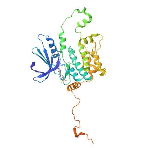
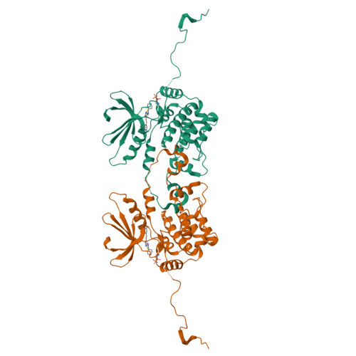
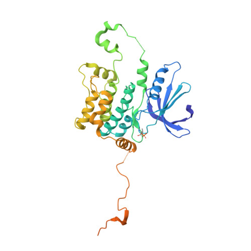
    Germinal-center kinase-like kinase (GLK, Map4k3), a GCK-I family kinase, plays multiple roles in regulating apoptosis, amino acid sensing, and immune signaling. We describe here the crystal structure of an activation loop mutant of GLK kinase domain bound to an inhibitor. The structure reveals a weakly associated, activation-loop swapped dimer with more than 20 amino acids of ordered density at the carboxy-terminus. This C-terminal PEST region binds intermolecularly to the hydrophobic groove of the N-terminal domain of a neighboring molecule. Although the GLK activation loop mutant crystallized demonstrates reduced kinase activity, its structure demonstrates all the hallmarks of an "active" kinase, including the salt bridge between the C-helix glutamate and the catalytic lysine. Our compound displacement data suggests that the effect of the Ser170Ala mutation in reducing kinase activity is likely due to its effect in reducing substrate peptide binding affinity rather than reducing ATP binding or ATP turnover. This report details the first structure of GLK; comparison of its activation loop sequence and P-loop structure to that of Map4k4 suggests ideas for designing inhibitors that can distinguish between these family members to achieve selective pharmacological inhibitors.


  • Organizational Affiliation

    Department of Drug Discovery, Biogen Inc., 115 Binney Street, Cambridge, MA, 02142.


Macromolecules
Find similar proteins by:  (by identity cutoff)  |  3D Structure
Entity ID: 1
MoleculeChains Sequence LengthOrganismDetailsImage
Mitogen-activated protein kinase kinase kinase kinase 3370Homo sapiensMutation(s): 1 
Gene Names: MAP4K3RAB8IPL1
EC: 2.7.11.1
UniProt & NIH Common Fund Data Resources
Find proteins for Q8IVH8 (Homo sapiens)
Explore Q8IVH8 
Go to UniProtKB:  Q8IVH8
PHAROS:  Q8IVH8
GTEx:  ENSG00000011566 
Entity Groups  
Sequence Clusters30% Identity50% Identity70% Identity90% Identity95% Identity100% Identity
UniProt GroupQ8IVH8
Sequence Annotations
Expand
  • Reference Sequence
Small Molecules
Ligands 1 Unique
IDChains Name / Formula / InChI Key2D Diagram3D Interactions
6G2
Query on 6G2

Download Ideal Coordinates CCD File 
B [auth A]5-[2-(piperidin-4-yl)-1,3-thiazol-5-yl]-3-[(pyridin-4-yl)methoxy]pyridin-2-amine
C19 H21 N5 O S
HSEGDFMLRPWOHH-UHFFFAOYSA-N
Modified Residues  1 Unique
IDChains TypeFormula2D DiagramParent
TPO
Query on TPO
A
L-PEPTIDE LINKINGC4 H10 N O6 PTHR
Experimental Data & Validation

Experimental Data

  • Method: X-RAY DIFFRACTION
  • Resolution: 2.85 Å
  • R-Value Free: 0.206 
  • R-Value Work: 0.175 
  • R-Value Observed: 0.177 
  • Space Group: C 1 2 1
Unit Cell:
Length ( Å )Angle ( ˚ )
a = 92.703α = 90
b = 63.324β = 116.04
c = 64.916γ = 90
Software Package:
Software NamePurpose
PHENIXrefinement
XDSdata reduction
SCALAdata scaling
MOLREPphasing

Structure Validation

View Full Validation Report


Ligand Structure Quality Assessment 


Entry History 

Deposition Data

Revision History  (Full details and data files)

  • Version 1.0: 2016-10-26
    Type: Initial release
  • Version 1.1: 2016-11-02
    Changes: Database references
  • Version 1.2: 2017-02-08
    Changes: Database references
  • Version 1.3: 2024-04-03
    Changes: Data collection, Database references, Refinement description
  • Version 1.4: 2024-10-30
    Changes: Structure summary