5J56 | pdb_00005j56

RTA-V1C7


Experimental Data Snapshot

  • Method: X-RAY DIFFRACTION
  • Resolution: 1.80 Å
  • R-Value Free: 
    0.210 (Depositor), 0.216 (DCC) 
  • R-Value Work: 
    0.173 (Depositor), 0.182 (DCC) 
  • R-Value Observed: 
    0.175 (Depositor) 

wwPDB Validation 3D Report Full Report

Validation slider image for 5J56

This is version 1.6 of the entry. See complete history

Literature

Structural Analysis of Single Domain Antibodies Bound to a Second Neutralizing Hot Spot on Ricin Toxin's Enzymatic Subunit.

Rudolph, M.J.Vance, D.J.Cassidy, M.S.Rong, Y.Mantis, N.J.

(2017) J Biological Chem 292: 872-883

  • DOI: https://doi.org/10.1074/jbc.M116.758102
  • Primary Citation Related Structures: 
    5BOZ, 5J56, 5J57

  • PubMed Abstract: 

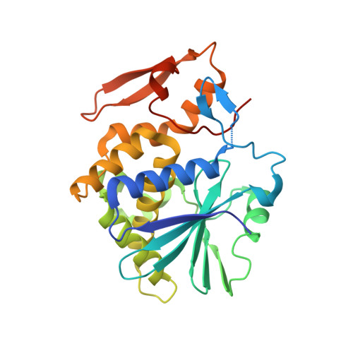
    Ricin toxin is a heterodimer consisting of RTA, a ribosome-inactivating protein, and RTB, a lectin that facilitates receptor-mediated uptake into mammalian cells. In previous studies, we demonstrated that toxin-neutralizing antibodies target four spatially distinct hot spots on RTA, which we refer to as epitope clusters I-IV. In this report, we identified and characterized three single domain camelid antibodies (V H H) against cluster II. One of these V H Hs, V5E1, ranks as one of the most potent ricin-neutralizing antibodies described to date. We solved the X-ray crystal structures of each of the three V H Hs (E1, V1C7, and V5E1) in complex with RTA. V5E1 buries a total of 1,133 Å 2 of surface area on RTA and makes primary contacts with α-helix A (residues 18-32), α-helix F (182-194), as well as the F-G loop. V5E1, by virtue of complementarity determining region 3 (CDR3), may also engage with RTB and potentially interfere with the high affinity galactose-recognition element that plays a critical role in toxin attachment to cell surfaces and intracellular trafficking. The two other V H Hs, E1 and V1C7, bind epitopes adjacent to V5E1 but display only weak toxin neutralizing activity, thereby providing structural insights into specific residues within cluster II that may be critical contact points for toxin inactivation.


  • Organizational Affiliation
    • From the New York Structural Biology Center, New York, New York 10027, mrudolph@nysbc.org.

Macromolecule Content 

  • Total Structure Weight: 44.53 kDa 
  • Atom Count: 3,262 
  • Modeled Residue Count: 383 
  • Deposited Residue Count: 397 
  • Unique protein chains: 2

Macromolecules

Find similar proteins by:|  3D Structure
Entity ID: 1
MoleculeChains  Sequence LengthOrganismDetailsImage
Ricin268Ricinus communisMutation(s): 0 
EC: 3.2.2.22
UniProt
Find proteins for P02879 (Ricinus communis)
Explore P02879 
Go to UniProtKB:  P02879
Entity Groups
Sequence Clusters30% Identity50% Identity70% Identity90% Identity95% Identity100% Identity
UniProt GroupP02879
Sequence Annotations
Expand
Reference Sequence
Find similar proteins by:|  3D Structure
Entity ID: 2
MoleculeChains  Sequence LengthOrganismDetailsImage
VHH single chain antibody V1C7129Vicugna pacosMutation(s): 0 

Small Molecules

Ligands 4 Unique
IDChains Name / Formula / InChI Key2D Diagram3D Interactions
PG4

Query on PG4


Download:Ideal Coordinates CCD File
C [auth A]TETRAETHYLENE GLYCOL
C8 H18 O5
UWHCKJMYHZGTIT-UHFFFAOYSA-N
EDO

Query on EDO


Download:Ideal Coordinates CCD File
F [auth A]1,2-ETHANEDIOL
C2 H6 O2
LYCAIKOWRPUZTN-UHFFFAOYSA-N
ACY

Query on ACY


Download:Ideal Coordinates CCD File
D [auth A],
E [auth A]
ACETIC ACID
C2 H4 O2
QTBSBXVTEAMEQO-UHFFFAOYSA-N
CL

Query on CL


Download:Ideal Coordinates CCD File
G [auth A],
H [auth A],
I [auth A]
CHLORIDE ION
Cl
VEXZGXHMUGYJMC-UHFFFAOYSA-M

Experimental Data & Validation

Experimental Data

  • Method: X-RAY DIFFRACTION
  • Resolution: 1.80 Å
  • R-Value Free:  0.210 (Depositor), 0.216 (DCC) 
  • R-Value Work:  0.173 (Depositor), 0.182 (DCC) 
  • R-Value Observed: 0.175 (Depositor) 
Space Group: P 32 2 1
Unit Cell:
Length ( Å )Angle ( ˚ )
a = 64.768α = 90
b = 64.768β = 90
c = 215.016γ = 120
Software Package:
Software NamePurpose
SCALEPACKdata scaling
PHASERphasing
PHENIXrefinement
PDB_EXTRACTdata extraction
HKL-2000data reduction

Structure Validation

View Full Validation Report


Entry History 

& Funding Information

Deposition Data


Funding OrganizationLocationGrant Number
National Institutes of Health/National Institute Of Allergy and Infectious Diseases (NIH/NIAID)United StatesHHSN272201400021C

Revision History  (Full details and data files)

  • Version 1.0: 2016-12-07
    Type: Initial release
  • Version 1.1: 2016-12-21
    Changes: Database references
  • Version 1.2: 2017-02-01
    Changes: Database references
  • Version 1.3: 2017-04-12
    Changes: Source and taxonomy, Structure summary
  • Version 1.4: 2017-09-13
    Changes: Author supporting evidence
  • Version 1.5: 2019-12-11
    Changes: Author supporting evidence, Derived calculations
  • Version 1.6: 2024-03-06
    Changes: Data collection, Database references