5J4V | pdb_00005j4v

The crystal structure of Inhibitor Bound to JCV Helicase


Experimental Data Snapshot

  • Method: X-RAY DIFFRACTION
  • Resolution: 2.94 Å
  • R-Value Free: 
    0.247 (Depositor), 0.253 (DCC) 
  • R-Value Work: 
    0.165 (Depositor), 0.177 (DCC) 
  • R-Value Observed: 
    0.169 (Depositor) 

wwPDB Validation 3D Report Full Report

Validation slider image for 5J4V

Ligand Structure Quality Assessment 


This is version 1.2 of the entry. See complete history

Literature

Fragment-Based Discovery of Dual JC Virus and BK Virus Helicase Inhibitors.

Bonafoux, D.Nanthakumar, S.Bandarage, U.K.Memmott, C.Lowe, D.Aronov, A.M.Bhisetti, G.R.Bonanno, K.C.Coll, J.Leeman, J.Lepre, C.A.Lu, F.Perola, E.Rijnbrand, R.Taylor, W.P.Wilson, D.Zhou, Y.Zwahlen, J.Ter Haar, E.

(2016) J Med Chem 59: 7138-7151

  • DOI: https://doi.org/10.1021/acs.jmedchem.6b00486
  • Primary Citation Related Structures: 
    5J40, 5J47, 5J4V, 5J4Y

  • PubMed Abstract: 

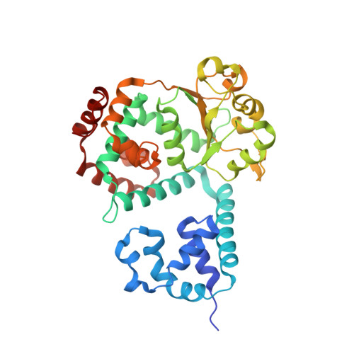
    There are currently no treatments for life-threatening infections caused by human polyomaviruses JCV and BKV. We therefore report herein the first crystal structure of the hexameric helicase of JCV large T antigen (apo) and its use to drive the structure-based design of dual JCV and BKV ATP-competitive inhibitors. The crystal structures obtained by soaking our early inhibitors into the JCV helicase allowed us to rapidly improve the biochemical activity of our inhibitors from 18 μM for the early 6-(2-methoxyphenyl)- and the 6-(2-ethoxyphenyl)-[1,2,4]triazolo[3,4-b][1,3,4]thiadiazole hits 1a and 1b to 0.6 μM for triazolopyridine 12i. In addition, we were able to demonstrate measurable antiviral activity in Vero cells for our thiazolopyridine series in the absence of marked cytotoxicity, thus confirming the usefulness of this approach.


  • Organizational Affiliation
    • Vertex Pharmaceuticals, Incorporated , 50 Northern Avenue, Boston, Massachusetts 02210, United States.

Macromolecule Content 

  • Total Structure Weight: 43.05 kDa 
  • Atom Count: 2,930 
  • Modeled Residue Count: 358 
  • Deposited Residue Count: 372 
  • Unique protein chains: 1

Macromolecules

Find similar proteins by:|  3D Structure
Entity ID: 1
MoleculeChains  Sequence LengthOrganismDetailsImage
Large T antigen372JC polyomavirusMutation(s): 13 
EC: 3.6.4 (PDB Primary Data), 5.6.2.4 (UniProt)
UniProt
Find proteins for P03072 (JC polyomavirus)
Explore P03072 
Go to UniProtKB:  P03072
Entity Groups
Sequence Clusters30% Identity50% Identity70% Identity90% Identity95% Identity100% Identity
UniProt GroupP03072
Sequence Annotations
Expand
Reference Sequence

Experimental Data & Validation

Experimental Data

  • Method: X-RAY DIFFRACTION
  • Resolution: 2.94 Å
  • R-Value Free:  0.247 (Depositor), 0.253 (DCC) 
  • R-Value Work:  0.165 (Depositor), 0.177 (DCC) 
  • R-Value Observed: 0.169 (Depositor) 
Space Group: P 6
Unit Cell:
Length ( Å )Angle ( ˚ )
a = 109.92α = 90
b = 109.92β = 90
c = 66.86γ = 120
Software Package:
Software NamePurpose
Aimlessdata scaling
BUSTERrefinement
PDB_EXTRACTdata extraction

Structure Validation

View Full Validation Report


Ligand Structure Quality Assessment 


Entry History 

Deposition Data

  • Released Date: 2016-07-20 
  • Deposition Author(s): Ter Haar, E.

Revision History  (Full details and data files)

  • Version 1.0: 2016-07-20
    Type: Initial release
  • Version 1.1: 2016-08-24
    Changes: Database references
  • Version 1.2: 2024-03-06
    Changes: Data collection, Database references, Derived calculations