5J34 | pdb_00005j34

Isopropylmalate dehydrogenase K232M mutant


Experimental Data Snapshot

  • Method: X-RAY DIFFRACTION
  • Resolution: 1.83 Å
  • R-Value Free: 
    0.185 (Depositor), 0.190 (DCC) 
  • R-Value Work: 
    0.165 (Depositor), 0.174 (DCC) 
  • R-Value Observed: 
    0.166 (Depositor) 

Starting Model: experimental
View more details

wwPDB Validation 3D Report Full Report

Validation slider image for 5J34

This is version 1.4 of the entry. See complete history

Literature

Structure and Mechanism of Isopropylmalate Dehydrogenase from Arabidopsis thaliana: INSIGHTS ON LEUCINE AND ALIPHATIC GLUCOSINOLATE BIOSYNTHESIS.

Lee, S.G.Nwumeh, R.Jez, J.M.

(2016) J Biological Chem 291: 13421-13430

  • DOI: https://doi.org/10.1074/jbc.M116.730358
  • Primary Citation Related Structures: 
    5J32, 5J33, 5J34

  • PubMed Abstract: 

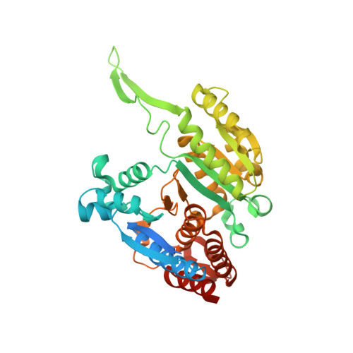
    Isopropylmalate dehydrogenase (IPMDH) and 3-(2'-methylthio)ethylmalate dehydrogenase catalyze the oxidative decarboxylation of different β-hydroxyacids in the leucine- and methionine-derived glucosinolate biosynthesis pathways, respectively, in plants. Evolution of the glucosinolate biosynthetic enzyme from IPMDH results from a single amino acid substitution that alters substrate specificity. Here, we present the x-ray crystal structures of Arabidopsis thaliana IPMDH2 (AtIPMDH2) in complex with either isopropylmalate and Mg(2+) or NAD(+) These structures reveal conformational changes that occur upon ligand binding and provide insight on the active site of the enzyme. The x-ray structures and kinetic analysis of site-directed mutants are consistent with a chemical mechanism in which Lys-232 activates a water molecule for catalysis. Structural analysis of the AtIPMDH2 K232M mutant and isothermal titration calorimetry supports a key role of Lys-232 in the reaction mechanism. This study suggests that IPMDH-like enzymes in both leucine and glucosinolate biosynthesis pathways use a common mechanism and that members of the β-hydroxyacid reductive decarboxylase family employ different active site features for similar reactions.


  • Organizational Affiliation
    • From the Department of Biology, Washington University, St. Louis, Missouri 63130.

Macromolecule Content 

  • Total Structure Weight: 174.36 kDa 
  • Atom Count: 12,547 
  • Modeled Residue Count: 1,436 
  • Deposited Residue Count: 1,620 
  • Unique protein chains: 1

Macromolecules

Find similar proteins by:|  3D Structure
Entity ID: 1
MoleculeChains  Sequence LengthOrganismDetailsImage
3-isopropylmalate dehydrogenase 2, chloroplastic
A, B, C, D
405Arabidopsis thalianaMutation(s): 1 
Gene Names: IMDH2IMDHAt1g80560T21F11.11
EC: 1.1.1.85
UniProt
Find proteins for P93832 (Arabidopsis thaliana)
Explore P93832 
Go to UniProtKB:  P93832
Entity Groups
Sequence Clusters30% Identity50% Identity70% Identity90% Identity95% Identity100% Identity
UniProt GroupP93832
Sequence Annotations
Expand
Reference Sequence

Small Molecules

Ligands 2 Unique
IDChains Name / Formula / InChI Key2D Diagram3D Interactions
SO4

Query on SO4


Download:Ideal Coordinates CCD File
F [auth A]
G [auth A]
H [auth A]
J [auth B]
L [auth C]
F [auth A],
G [auth A],
H [auth A],
J [auth B],
L [auth C],
N [auth D]
SULFATE ION
O4 S
QAOWNCQODCNURD-UHFFFAOYSA-L
MG

Query on MG


Download:Ideal Coordinates CCD File
E [auth A],
I [auth B],
K [auth C],
M [auth D]
MAGNESIUM ION
Mg
JLVVSXFLKOJNIY-UHFFFAOYSA-N

Experimental Data & Validation

Experimental Data

  • Method: X-RAY DIFFRACTION
  • Resolution: 1.83 Å
  • R-Value Free:  0.185 (Depositor), 0.190 (DCC) 
  • R-Value Work:  0.165 (Depositor), 0.174 (DCC) 
  • R-Value Observed: 0.166 (Depositor) 
Space Group: P 1 21 1
Unit Cell:
Length ( Å )Angle ( ˚ )
a = 76.803α = 90
b = 76.82β = 90.22
c = 209.497γ = 90
Software Package:
Software NamePurpose
PHENIXrefinement
HKL-3000data reduction
HKL-3000data scaling
PHASERphasing

Structure Validation

View Full Validation Report


Entry History 

Deposition Data

Revision History  (Full details and data files)

  • Version 1.0: 2016-05-11
    Type: Initial release
  • Version 1.1: 2016-05-18
    Changes: Database references
  • Version 1.2: 2016-07-06
    Changes: Database references
  • Version 1.3: 2024-03-06
    Changes: Data collection, Database references, Derived calculations
  • Version 1.4: 2024-04-03
    Changes: Refinement description